|
Entomotropica
antes/formerly Boletín de Entomología Venezolana
Vol. 17(3): 225-294. Diciembre 2002
|
ISSN 1317-5262
|
Revision of the Neotropical genus Trizogeniates Ohaus (Coleoptera: Scarabaeidae: Rutelinae: Geniatini)
Karla Villatoro
University of Nebraska State Museum, Division of Entomology, W 436 Nebraska Hall, Lincoln, NE 68588-0514, U.S.A. E-mail: [email protected]
Recibido: 30-viii-2001
Aceptado: 21-x1-2001
Correcciones devueltas por el autor: 04-vi-2002
Abstract
Villatoro K 2002. Revision of the Neotropical genus Trizogeniates Ohaus (Coleoptera: Scarabaeidae: Rutelinae: Geniatini). Entomotropica 17(3):225-294.
A comprehensive, systematic treatment of the poorly studied Neotropical genus Trizogeniates (Coleoptera: Scarabaeidae: Rutelinae: Geniatini) is provided. This revision includes a key to species, species descriptions, and distributional and temporal data. Based on this study, the genus Trizogeniates now includes 30 species. The following eight species are new: Trizogeniates ohausi Villatoro, T. venezuelensis Villatoro, T. geminatus Villatoro, T. caiporae Villatoro, T. crispospinatus Villatoro, T. catsus Villatoro, T. aphilus Villatoro, and T. eris Villatoro. Trizogeniates zischkai Martínez is a new synonym of T. temporalis Ohaus; T. grandis Ohaus is a new synonym of T. cribicollis (Lucas). T. andicola Ohaus is a new synonym of T. tibialis Ohaus; and T. navajasi Martínez is a new synonym of T. terricola Ohaus. T. vittatus subandinus Martínez is a new synonym of T. vittatus Ohaus. Bolax vittata Casey is a new synonym of T. foveicollis Ohaus and a secondary junior homonym of T. vittatus (Lucas). T. caseyi Villatoro is created as a replacement name for T. vittatus (Casey). Neotypes and lectotypes are designated and discussed in the appropriate cases.
Additional key words: Bearded scarab beetle, South America, systematics, taxonomy.
Resumen
Villatoro K. 2002. Revisión del género neotropical Trizogeniates Ohaus (Coleoptera: Scarabaeidae: Rutelinae: Geniatini). Entomotropica 17(2):225-294.
Se presenta un estudio sistemático del poco conocido género neotropical Trizogeniates (Coleoptera: Scarabaeidae: Rutelinae: Geniatini). Esta revisión incluye una clave ilustrada para especies, descripciones para cada especie y datos sobre su distribución temporal y geográfica. Basándose en este trabajo, el género Trizogeniates ahora incluye 30 especies. Las siguientes ocho especies son nuevas: Trizogeniates ohausi Villatoro, T. venezuelensis Villatoro, T. geminatus Villatoro, T. caiporae Villatoro, T. crispospinatus Villatoro, T. catsus Villatoro, T. aphilus Villatoro y T. eris Villatoro. Trizogeniates zischkai Martínez es un nuevo sinónimo de T. temporalis Ohaus y T. grandis Ohaus es un nuevo sinónimo de T. cribicollis (Lucas). T. andicola Ohaus es un nuevo sinónimo de T. tibialis Ohaus. T. navajasi Martínez es un nuevo sinónimo de T. terricola Ohaus. T. vittatus subandinus Martínez es un nuevo sinónimo de T. vittatus Ohaus. Bolax vittata Casey es un nuevo sinónimo de T. foveicollis Ohaus y un homonimo junior secundario de T. vittatus (Lucas). El nombre T. caseyi Villatoro es creado como remplazo de T. vittatus (Casey). Neotipos y lectotipos son designados y se discuten en los casos apropiados.
Palabras clave adicionales: Escarabajo, sistemática, Suramérica, taxonomía.
Introduction
The genus Trizogeniates is a group of scarabs in the subfamily Rutelinae. These scarabs are moderately-sized (about 2 cm), and their coloration ranges from black to tawny; several species exhibit blackish elytral vittae alternating with tawny vittae. Members of this genus can be easily distinguished by the presence of a unique stridulatory apparatus that consists of a passive structure (stridulatory ridge) and an active structure (stridulatory file). The ridge is setose and located along the epipleuron from the metacoxa to the elytral apex (Figure 36). The active structure consists of numerous, fine ridges giving it the functionality of a file and has the appearance of a matte spot found on the dorsolateral apex of the metafemur (Figure 35). Stridulation is produced by rubbing the file against the ridge of setae. This stridulatory apparatus was first described in 1903 by Ohaus, but it was not until 1917 that he decided to create the genus Trizogeniates based on this character. I consider Trizogeniates to be a monophyletic group since the apparatus is a unique, derived character shared by all the species in the genus.
Trizogeniates, like most other genera in the Geniatini, is a poorly known group. A key or treatment of the genus had never been done. The majority of scientific papers on Trizogeniates have been new species descriptions. The only publications dealing with the genus as a whole are the Coleopterorum Catalogus (Machatschke 1972, 1974), the Genera Insectorum (Machatschke 1965), and Blackwelder’s (1944) catalog. The lack of knowledge about Trizogeniates has resulted in a perpetuation of misidentifications, erroneous redescriptions of described species, and a backlog of unidentified specimens in museum collections.
This research provides a detailed study of the species of Trizogeniates and a means to identify specimens with taxonomic keys, illustrations, and detailed descriptions of all species. The objectives of this study are: 1) characterize each species of Trizogeniates based on morphological characters, and 2) provide keys, descriptions, diagnoses, illustrations, and temporal and geographic distributions for the species. This study provides the foundation for further biological and evolutionary studies in the Geniatini.
Friedrich Ohaus and Antonio Martínez were the two major contributors to the genus, describing almost 80% of the species (52% and 14%, respectively). Taxonomic decisions, like neotype, incertae sedis, and synonymy designations were necessary tasks due to the following problems: 1) Species descriptions are not detailed enough to separate species. Many species descriptions are general and superficial and used characters that apply to many species of Trizogeniates. Ohaus’ descriptions lack key characters like shape of the parameres, shape of prosternal shield, characters of the last sternite and pygidium, and shape of the epipleuron. Although some of his characters are diagnostic (e.g., curvature on apex of the metatibial spur, length of the first metatarsomere, sculpture of the pygidium), they are not used consistently throughout his descriptions and are not enough to distinguish similar species; 2) Martínez did not examine Ohaus types and so he created new species that were previously described by Ohaus; and 3) Type specimens were lost. A total of 35% of the types specimens were not found. In some cases, identifications of species by the authors themselves were not reliable. For example, I found several series of specimens identified by Ohaus as one species, when it was actually composed of two or more species. Without a thorough description, a type specimen, or reliable species identifications, some species remain of uncertain identity.
Natural history and larvae are unknown for all of the species, but I know that they are attracted to lights at night. Species of Trizogeniates inhabit semideciduous forests and cloud forest (sea level-2400 m) from Costa Rica to northern Argentina. Brazil is high in diversity and has more than half of the species.
Materials and Methods
Taxonomic Material
A total of 1000 specimens were used for the revision of the genus Trizogeniates. Specimens examined for this study, including type specimens, provided by 42 institutions and private collections. Acronyms for loaning institutions follow the on-line version of Insect and Spider Collections of the World (Evenhuis et al. 2000).
|
AMNH
|
American Museum of Natural History, New York, NY.
|
|
ANSP
|
Academy of Natural Sciences, Philadelphia, PA.
|
|
BCRC
|
Brett C. Ratcliffe Collection, Lincoln, NE.
|
|
BMNH
|
The Natural History Museum, London, United Kingdom.
|
|
CASC
|
California Academy of Sciences, San Francisco, CA.
|
|
CMNC
|
Canadian Museum of Nature, Ottawa, Canada.
|
|
CMNH
|
Carnegie Museum of Natural History, Pittsburgh, PA.
|
|
CNCI
|
Canadian National Collection of Insects, Ottawa, Canada.
|
|
DCCC
|
David C. Carlson Collection, Orangevale, CA.
|
|
DEES
|
Universidade de Săo Paulo, Săo Paulo, Brazil.
|
|
DEI
|
Deutsches Entomologisches Institut, Eberswalde Finow, Germany.
|
|
DJCCC
|
Daniel J. Curoe Collection, Palo Alto, CA.
|
|
EGRC
|
Edward G. Riley Collection, College Station, TX.
|
|
FMNH
|
Field Museum of Natural History, Chicago, IL.
|
|
FSCA
|
Florida State Collection of Arthropods, Gainsville, FL.
|
|
HAHC/CMNC
|
Henry and Anne Howden Collection, (currently housed at CMNC and includes Antonio Martínez Collection).
|
|
INBC
|
Instituto Nacional de Biodiversidad (INBio), Santo Domingo de Heredia, Costa Rica.
|
|
JEWC
|
James E. Wappes Collection, Bulverde, TX.
|
|
KSUC
|
Kansas State University Collection, Manhattan, KS.
|
|
LACM
|
Museum of Natural History, Los Angeles, CA.
|
|
MACN
|
Museo Argentino de Ciencias Naturales, Buenos Aires, Argentina.
|
|
MCZC
|
Museum of Comparative Zoology, Cambridge, MA.
|
|
MIZA
|
Museo del Instituto de Zoología Agrícola, Maracay, Venezuela.
|
|
MLJC
|
Mary Liz Jameson Collection, Lincoln, NE.
|
|
MLP
|
Museo de La Plata, Universidad Nacional de La Plata, La Plata, Argentina.
|
|
MLUH
|
Wissenschaftsbereich Zoologie von Martin-Luther-Universität, Halle, Germany (currently housing Burmeister Collection).
|
|
MNHN
|
Muséum National d'Histoire Naturelle, Paris, France.
|
|
NHMB
|
Naturhistorisches Museum, Basel, Switzerland (currently housing G. Frey Collection).
|
|
QCAZ
|
Entomology Museum, Pontificia Universidad Católica del Ecuador, Quito, Ecuador.
|
|
RFMC
|
Roy F. Morris II Collection, Lakeland, FL.
|
|
ROME
|
Royal Ontario Museum, Ontario, Canada.
|
|
SEMC
|
Snow Entomological Museum, Lawrence, KS.
|
|
SMTD
|
Staatliches Museum für Tierkunde, Dresden, Germany.
|
|
TAMU
|
Texas A & M University, College Station, TX.
|
|
UASC
|
Museo de Historia Natural “Noel Kempff Mercado”, Santa Cruz, Bolivia.
|
|
UMRM
|
W. R. Enns Entomology Museum, Columbia, MO.
|
|
UNSM
|
University of Nebraska State Museum, Lincoln, NE.
|
|
USNM
|
U. S. National Museum of Natural History, Washington, D.C. (currently housed at University of Nebraska).
|
|
VMCC
|
Vladislav Maly Collection, Prague, Czech Republic.
|
|
WBWC
|
William B. Warner Collection, Chandler, AZ.
|
|
ZMHB
|
Museum für Naturkunde der Humboldt-Universität, Berlin, Germany.
|
|
ZSMC
|
Zoologische Staatssammlung München, Munich, Germany.
|
Designation of Lectotypes and Neotypes
The International Code of Zoological Nomenclature (CZN 1999) requires that designations of lectotypes after 1999 must “contain an express statement of the taxonomic purpose of the designation” (74.7.3). In this work lectotypes were designated for several species in order to preserve the nomenclatural stability by selecting one specimen as the sole name bearing type of the taxon. The lectotype specimen serves to tie the published name to an actual specimen and as a reference standard for the taxon. Label data associated with the lectotype specimens and institution where specimens were deposited are included under the description of each species. Lectotype specimens were designated for the following species names: T. bicolor Ohaus, T. catoxanthus Ohaus, T. dispar (Burmeister), T. montanus Ohaus, T. grandis Ohaus, T. planipennis Ohaus, T. temporalis Ohaus, and T. terricola Ohaus. Ohaus did not designate holotypes within his type series, but instead he placed “type” and/or “cotype” labels on specimens.
In Ohaus’ descriptions of Trizogeniates species, he did not indicate the exact number of specimens in the type series. The only information provided were ranges of measurements that indicate that the type series included more than one specimen, and he also indicated if the type series had both or only one sex.
Ohaus also placed his type or cotype labels on specimens after the species description was originally published (Jameson 1998). These types were invalidly designated by Ohaus. They were identified based on incorrect sex of specimen or locality data that did not agree with the original description. For those specimens, my “invalid type” label was placed under the specimen. Invalid type designations are also discussed in the appropriate species under the “Remarks” section.
The International Code of Zoological Nomenclature (CZN 1999) requires that a neotype “is validly designated when there is an exceptional need and only when that need is stated expressly” (75.3). In this work, four neotypes were designated to preserve the stability of nomenclature by selecting one specimen as the sole name-bearing type of the taxon when the original name-bearing type specimen(s) were lost or destroyed. The neotype specimen serves to tie the published name to an actual specimen and as a reference standard for the taxon. Neotypes specimens were designated for the following species names: T. foveicollis Ohaus, T. laticollis Ohaus, T. tibialis Ohaus, and T. trivittatus Ohaus. Neotypes for these names are necessary due to the long history of taxonomic neglect in this genus. Label data and institution where the types are stored were included within the descriptions.
Dissection
In this study, dissection of specimens was necessary for the examination of mouthparts and genitalia. The dissection technique followed that of Jameson (1998). Dried specimens were softened by boiling them in distilled water for several minutes (with a drop of detergent to break up fat). Mouthparts (mentum, maxilla, mandible, and sometimes labrum) were extracted using microforceps and insect pins. The aedeagus was extracted using one of the following techniques (depending on the condition of the specimen): 1) The aedeagus was extracted through the genital opening. Microforceps and insect pins were sufficient instruments for extracting the genitalia. 2) If genitalia were difficult to extract through the genital opening, they were dissected by carefully removing the abdomen at the juncture between the metathorax and the first abdominal sternite. Mouthparts and parameres were card-mounted using ethylose glue and then pinned beneath the specimen. In most cases, the right mandible, right maxilla, and mentum were extracted, thus leaving the left side intact. Following the examination of all characters, the abdomen was replaced (using ethylose glue).
Species Concept
The phylogenetic species concept (Wheeler and Platnick 2000) was applied in this work: “A species is the smallest aggregation of (sexual) populations or (asexual) lineages diagnosable by a unique combination of character states”.
Character Examination
Descriptions and keys were constructed using internal as well as external characters. These structures were examined with a dissecting microscope (6.3 to 50X power) and fiber-optic lights. Measurements were taken with the ocular micrometer of the microscope. As many characters as possible were used in this study, but only those characters that proved to have low intraspecific variability were chosen to characterize species. Characters that did not require dissection were favored for species descriptions. Some characters historically used in species descriptions proved to vary considerably. These characters are discussed but were not used to separate species.
Species in the genus Trizogeniates are characterized by a combination of several character states. Some character states are autapomorphic for species (e.g., form of the pronotum or shape of the labrum), and some characters states are synapomorphic for several species, suggesting species groups within the genus.
The following characters were found to be taxonomically useful:
Body measurements. Length was measured from the apex of the pronotum to the apex of the elytra. Body width was measured across the elytral humeri.
Elytral intervals. Each elytron has 10 intervals that were numbered I-X from the elytral suture to the elytral margin (Figure 29). Intervals were described based on shape, distribution of ocellate punctures, and color. The shape of the intervals was defined as flat, weakly convex, or strongly convex. Ocellate punctures are present only in even intervals, and they can be distributed evenly over the interval or in a longitudinal row. Color pattern was defined as vittate or non-vittate. Vittate species are those that have the even intervals darker than the odd intervals. Non-vittate species are those that are monochromatic (or where darker coloration occurs along the suture and at the apices).
Puncture density. Punctures were considered dense if they were nearly confluent to less than two puncture diameters apart, moderately dense if punctures were from two to six puncture diameters apart, and sparse if punctures were separated by more than six puncture diameters.
Puncture size. Punctures are defined as small when 0.02 mm or smaller; moderate when 0.02-0.07, moderately large when 0.07-0.12, and large when 0.12 or larger. Punctures were ocellate or simple, round or oval-shaped, and shallow or deep.
Pilosity. Metatarsomeres and the prosternal shield exhibited variability in the density of setae. Setae were defined as dense if the surface was not visible through the setae, moderately dense if the surface was visible but with many setae, and sparsely dense if there were few setae.
Interocular width. This measurement equals the number of transverse eye diameters that fit between the eyes.
Labrum. The labrum was measured along the middle from the base to the apex of the labral tooth. This measurement was used in comparison to the thickness of the clypeal apex (measured at the middle from top to bottom). There were two states for the width of the labrum: a) thick (medial length subequal to thickness of the clypeal apex, Figure 15) and b) thin (medial length less than half the thickness of the clypeal apex, Figure 14).
Maxillary teeth. Teeth were assigned a number from the outermost to innermost tooth: 1st tooth was the external tooth, 2nd tooth was the ventral tooth and is usually ventrally expanded, 3rd tooth was the dorsal tooth, 4th and 5th were the innermost teeth, usually located behind the second and third teeth (Figures 18-23).
Prosternal shield. The following four characters were diagnostic: 1) shape of apical margin (angulate or rounded), 2) disc convexity (weak or strong), 3) size (reduced or broad, Figures 59-60), and 4) pilosity (densely or sparsely setose, Figures 58-60).
Epipleuron. The width of the epipleuron was measured in two places: base of the metepisternum and the area laterad of the abdominal sternites. At the base of the metepisternum, the epipleuron width may be: 1) narrow (width of epipleuron at base of metepisternum shorter than width of base of metepisternum, Figure 30), 2) moderately broad (width of epipleuron at base of metepisternum equal to width of base of metepisternum), or 3) broad (width of epipleuron at base of metepisternum wider than width of base of metepisternum, Figure 31). In the area laterad of the abdominal sternites, the epipleural region laterad of the stridulatory ridge may be broad or narrow (Figures 37-38). This region also varied in shape. It may be flat (oblique or horizontal) or concave.
Stridulatory ridge. At the metacoxa, the stridulatory ridge originates: 1) near the inner edge of the epipleuron, 2) center of the epipleuron, or 3) near the outer edge of the epipleuron (Figures 32-34). The stridulatory ridge may become confluent with the marginal bead of the elytron or not. If it does become confluent, it may be confluent for its entire length or it may be confluent only to the region posterior to the metacoxa, to the 1st sternite, the 2nd sternite, or the 3rd sternite.
Last sternite. Males have an emargination at the apex of the last sternite that may be: 1) deep (middle of emargination more than 1/2 length of sternite) or 2) shallow (middle of emargination less than 1/2 length of sternite). The middle of the emargination may be rounded, angulate, or with a rounded notch (Figures 74-76). The apex of the last sternite of females may be: 1) simple, 2) crenulate, 3) emarginated, 4) emarginated with notch at the middle, 5) deeply bi-emarginate, or 6) shallowly bi-emarginate (Figures 77-82).
Metatibial spurs. In males, the outer spurs may be: 1) slightly curved and semicircular in cross section (Figure 44) or 2) strongly curved and circular in cross section (Figure 43). In males, the inner spurs may be: 1) slightly curved and semicircular in cross section (Figure 47), 2) strongly curved and circular in cross section (Figure 48), or 3) hooked at apex and circular in cross section (Figure 45-46, 49). In females, the apex of the inner spur may be rounded or truncate (Figure 50-51).
First sternite. The base of the first sternite at the middle may be simple, not ventrally produced (Figure 61) or ventrally produced (Figures 62-63).
Protarsomeres. Males possess expanded protarsomeres (except tarsomere 5) that vary in shape between species. Distinct shapes were defined by the relative length and the level of concavity in lateral view. Protarsomeres 2 and 3 may be: 1) elongate (widest width subequal to 2/3 length), 2) weakly elongate (widest width subequal to 5/6 length), or 3) stout (widest width subequal to length). The dorsal surface of protarsomeres 2 to 4 may be convex or flat. The length of protarsomere 5 can be: 1) short (length shorter than 1/2 length of protarsomeres 2-4), or 2) elongate (length longer than 1/2 length of protarsomeres 2-4).
Genitalia. Male genitalia were diagnostic for all species (Figures 83-113). The parameres may be symmetrical or not. Three species showed slight intraspecific variation. Female gonocoxites were not useful for separating species.
Genus Trizogeniates Ohaus
(Figures 1-113, Maps 1-6)
Trizogeniates Ohaus, 1917: 38.
Type species. Geniates vittatus Lucas, 1857: 134. Fixed by subsequent designation (Machatschke, 1965: 137).
Description. Scarabaeidae: Rutelinae: Geniatini. FORM (Figures 1-4): Elongate oval, sides subparallel, pygidium exposed, apex broadly rounded. HEAD: Surface punctate, more heavily sculptured in females. Frons without horns or concavities. Frontoclypeal suture complete, straight. Eye canthus simple, not carinate. Clypeus (Figures 5-9) with apex reflexed (in females more weakly reflexed), lacking bead. Mandibles (Figures 16-17) with baso-external edge round, apex with one reflexed, round tooth; inner teeth present or lacking. Labrum (Figures 14-15) apicomedially with forward-projecting tooth. Maxilla (Figures 18-23, 24) with baso-external edge of mala round, raised, with 3, 4, or 5 teeth; tooth 2 usually ventrally expanded, ventral margin rounded; other teeth smaller. Stipes of maxilla not produced. Mentum (Figures 12-13) in lateral view weakly to strongly convex. In ventral view, subrectangular; small sulcus present near insertion of palpus (extending to lateral margin), margin laterad of insertion with acute angle. Apex with median, dorsally-produced tooth; tooth not concave and sides not converging towards apex (except in T. goyanus). Disc sparsely setose. Antenna 10-segmented with 3-segmented club; club longer in males than in females. PRONOTUM (Figures 25-26): Widest at middle (except T. eris where widest at base); anterior angles acute (except T. eris). In frontal view, dorsal surface weakly convex. Surface variably punctate. Marginal bead complete. Scutellum with shape parabolic, apex weakly acute, length subequal to width. Surface variably punctate. ELYTRA (Figure 29): Width subequal to 3/4 length. Margin on sides and apex beaded; bead from base to metacoxae variably developed, after metacoxae well-developed. Surface with nine striae; striae variably impressed, longitudinal, punctate; punctures ocellate. Intervals 1-5 on disc, interval 6 on humerus, 7-10 laterad of humerus. Odd intervals weakly convex to strongly convex, punctate, punctures simple. Even intervals weakly convex to flat, punctate, punctures simple and ocellate; if even intervals darker than odd intervals, then ocellate punctures on entire interval; if even intervals not darker than odd intervals, ocellate punctures forming longitudinal row. Epipleuron (Figures 30-31) in cross section straight or angulate, not rounded. Ventral side of epipleuron from middle of metepisternum to apex with setose ridge; setae short to moderately long, thick. Elytral apex weakly rounded. Membrane visible from metacoxa to apex. PYGIDIUM (Figures 64-65): Shape subtriangular. Surface variably sculptured: smooth, punctate, rugose. Margin with sides and apex beaded. Apical bead simple, arcuate, biarcuate, or thickened at middle (Figures 66-71). VENTER: Prosternal shield present, shape variable, never protruding ventrally beyond apex of procoxae. Mesometasternal keel lacking. Mesosternum flat, not invaginated or strongly concave. In lateral view, male sternites flat, female sternites weakly convex. First sternite (Figures 61-63) with base produced or not produced ventrally. Last sternite (Figures 28, 29) of males with apical emargination and with row of setae; females with or without emargination. LEGS: Protibia with three teeth, basal tooth weakly removed from other teeth; inner apex with spur; base without protibial notch. Male protarsomeres 1-4 (Figure 28) dorsoventrally flattened, densely setose ventrally, setae short, tawny; 5th tarsomere cylindrical, lacking ventral pilosity; protarsal claw subequally split dorso-ventrally; unguitractor plate weakly exposed beyond apex of protarsomere 5, bisetose. Female protarsomeres 1-5 (Figure 27) subcylindrical (apex slightly wider than base), venter with sparse pilosity, setae moderately long, castaneous; protarsal claw subequally split dorso-ventrally; unguitractor plate weakly exposed beyond apex of protarsomere 5, bisetose. Meso- and metatibia each weakly expanded apically (mesotibial apex slightly more expanded in females); external edge with two weakly developed carinae; apex with spurs and spines, two spurs at inner apex placed in depression, spurs variable in shape. Surface variably punctate, metatibia more clearly sculptured than mesotibia. Male meso- and metatarsomeres 1-4 ventrally flattened, setose ventrally (mesotarsomeres less setose than protarsomeres, and metatarsomeres less setose than mesotarsomeres), setae short, tawny. Tarsal claw subequally split dorso-ventrally. Female meso- and metatarsomeres subcylindrical (apex slightly wider than base), venter with sparse pilosity; protarsal claw subequally split dorso-ventrally. Metafemur (Figure 35) dorsally with stridulatory file apicolaterally. Metatrochanter with apex produced or not beyond posterior border of femur. Metacoxa apex laterally square or rounded. HIND WING: Well developed hooks on precostal membrane present. Anterior edge from medial fold to apex of wing with seatae present. Vein AA1+2 less than one half length of vein AA3+4. PARAMERES (Figures 83-113): Diagnostic. FEMALE GONOCOXITES: Not diagnostic.
Diagnosis. Trizogeniates differs from other genera in the tribe by the following characters: metafemur (dorsal view) with stridulatory file apicolaterally and surface of epipleuron with setose, stridulatory ridge. Species of this genus could be confused with some species of Lobogeniates Ohaus or Geniates Ohaus because of the overall shape, appearance, and presence of blackish vittae. However, Trizogeniates can be separated from these genera by the following combination of character states: 1) stipes of maxilla not produced; 2) all tarsal claws subequally split dorso-ventrally; 3) disc of mentum lacking circular region of dense, ventrally produced setae. An electronic key to the genera of Geniatini is available at: http://www-museum.unl.edu/research/entomology/Guide/Rutelinae/Geniatini/GeniatiniK.htm
Distribution (Maps 1-6). Costa Rica to southern Brazil and northern Argentina. Found at elevations ranging from sea level to 2500 m.
Remarks. Some species were originally described in the genus Geniates and have since been transferred to Trizogeniates. Blackwelder (1944) incorrectly considered the genus to be feminine in gender, and he emended masculine names to feminine endings (Villatoro and Jameson 2000). A profile of the genus is available on-line at: http://www-museum.unl.edu/research/entomology/Guide/Rutelinae/Geniatini/Trizogeniates/Trizogeniates.htm.
Key to the species of Trizogeniates
(except T. apicalis, T. calcaratus, T. laevis, T. costatus, males of T. eris, and females of T. aphilus)
| 1 |
Protarsomeres dorsoventrally flattened, densely setose ventrally (Figure 28).
Males ....................................................................................................2
|
| |
|
| 1' |
Protarsomeres subcylindrical, not flattened, sparsely setose ventrally
(Figure 27). Females ...........................................................................27
|
| , |
|
| MALES |
|
| 2 |
Elytra with blackish vittae (Figures 3-4) .................................................3
|
| , |
|
| 2' |
Elytra without blackish vittae (Figures 1-2) ..........................................14
|
| , |
|
| 3 |
Apex of inner metatibial spur hooked (Figures 45-46, 49) .....................4
|
| , |
|
| 3' |
Apex of inner metatibial spur not hooked (Figures 47-48) .....................7
|
| , |
|
| 4 |
Length of protarsomere 2 longer than its widest width ...........................5 |
| , |
|
| 4' |
Length of protarsomere 2 equal to its widest width. Male genitalia as in
Figure 111 ............................................................T. trivittatus Ohaus
|
| , |
|
| 5 |
Apical emargination of last sternite without a rounded notch at middle
(Figures 74-75) ..................................................................................6
|
| , |
|
| 5' |
Apical emargination of last sternite with a rounded notch at middle
(Figure 76). Male genitalia as in Figure 108 ................T. tibialis Ohaus
|
| , |
|
| 6(3’) |
Apical emargination of last sternite almost V-shaped (Figure 75).
Male genitalia as in Figure 83 .......................T. aphilus Villatoro, n. sp.
|
| , |
|
| 6' |
Apical emargination of last sternite arcuate, not V-shaped (Figure 74).
Male genitalia as in Figures 91-92 ......................................................
.........................................................T. crispospinatus Villatoro n. sp.
|
| , |
|
| 7(3’) |
Inner metatibial spur weakly curved (Figure 47) and semicircular
in cross section ...................................................................................8
|
| , |
|
| 7’ |
Inner metatibial spur strongly curved (Figure 48) and transversally
rounded in cross section. Male genitalia as in Figure 105 ........................
............................................................................T. schmidti (Ohaus)
|
| , |
|
| 8 |
Disc of pygidium not rugose or pilose ..................................................9 |
| , |
|
| 8’ |
Disc of pygidium rugose and pilose (Figure 64). Male genitalia as
in Figure 84 ..........................................................T. barrerai Martínez
|
| , |
|
| 9 |
Clypeus subrectangular (Figure 6). Terminal segment of maxillary
palpus with broad, shagreened area (Figure 11). Male genitalia as
in Figure 104 ....................................................T. planipennis Ohaus
|
| , |
|
| 9’ |
Clypeus parabolic or trapezoidal (Figures 7-9). Terminal segment
of maxillary palpus with narrow, longitudinal, shagreened area
(Figure 10) ......................................................................................10
|
| , |
|
| 10 |
Epipleuron with base narrower or subequal to base of metepisternum
(Figure 30) ......................................................................................11
|
| , |
|
| 10’ |
Epipleuron with base wider than base of metepisternum (Figure 31).......
........................................................................................................12
|
| , |
|
| 11 |
Length of protarsomere 2 greater than its widest width. Male genitalia
as in Figure 89 ..............................................T. catsus Villatoro, n.sp.
|
| , |
|
| 11’ |
Length of protarsomere 2 shorter than its widest width. Male genitalia
as in Figure 94 ....................................................T. foveicollis Ohaus
|
| , |
|
| 12(10’) |
In anterior view, labrum with length subequal to length of reflexed
clypeal apex. Region next to clypeal tooth not concave (Figure 15)
........................................................................................................13
|
| , |
|
| 12’ |
In anterior view, labrum with length less than half length of reflexed
clypeal apex. Region next to clypeal tooth weakly concave (Figure 14).
Male genitalia as in Figure 96 ..................................T. goyanus Ohaus
|
| , |
|
| 13 |
Apex of first sternite at middle not produced ventrally (Figure 61).
Male genitalia as in Figure 113 ...............................T. vittatus (Lucas)
|
| , |
|
| 13’ |
Apex of first sternite at middle produced ventrally (Figure 63).
Male genitalia as in Figure 106 ............................T. temporalis Ohaus
|
| , |
|
| 14(2’) |
Metafemur with posterior margin at base lacking indentation (Figure 40)
........................................................................................................15
|
| , |
|
| 14’ |
Metafemur with posterior margin at base with indentation (Figure 39).
Male genitalia as in Figure 86 ...............................T. bordoni Martínez
|
| , |
|
| 15 |
Metafemur with posterior margin at apex weakly extended and weakly
rounded or angulate (Figure 42) .......................................................16
|
| , |
|
| 15’ |
Metafemur with posterior margin at apex strongly extended and rounded
(Figure 41). Male genitalia as in Figure 107 ............T. terricola Ohaus
|
| , |
|
| 16 |
Elytral region laterad of humerus declivous. Stridulatory ridge begins
at center of epipleuron or near outer edge of epipleuron (Figures 32-33)
.......................................................................................................17
|
| , |
|
| 16’ |
Elytral region laterad of humerus weakly declivous. Stridulatory ridge
begins at inner edge of epipleuron (Figure 34). Male genitalia as in
Figures 97-101 ....................................................T. laticollis Ohaus
|
| , |
|
| 17 |
Clypeus not subrectangular (Figures 7-9) ........................................18
|
| , |
|
| 17’ |
Clypeus subrectangular (Figure 5). Male genitalia as in Figure 90..........
.........................................................................T. cribicollis (Lucas)
|
| , |
|
| 18 |
Prosternal shield with long, sparse setae (Figures 59-60) ................19
|
| , |
|
| 18’ |
Prosternal shield with short, dense setae (Figure 58). Male genitalia
as in Figure 31h .................................................T. montanus Ohaus
|
| , |
|
| 19 |
Outer metatibial spur weakly curved, semicircular in cross section
along entire length, apex rounded (Figure 44) ..................................20
|
| , |
|
| 19’ |
Outer metatibial spur curved, circular in cross section along entire
length, apex pointed (Figure 43). Male genitalia as in Figure 85 ..........
...............................................................................T. bicolor Ohaus
|
| , |
|
| 20 |
Length of protarsomere 2 equal or subequal to its widest width .......21 |
| , |
|
| 20’ |
Length of protarsomere 2 greater than its widest width. Male
genitalia as in Figure 75 .......................T. geminatus Villatoro, n. sp.
|
| , |
|
| 21 |
Pygidium with disc rugopunctate, punctures horizontally elongate ........
.....................................................................................................22 |
| , |
|
| 21’ |
Pygidium with disc smooth, punctures simple (Figure 65). Male
genitalia as in Figure 110 ...............................T. travassosi Martínez
|
| , |
|
| 22 |
Metatarsomere 5 on ventral side with protuberance
(Figure 54-55, 57) ......................................................................23
|
| , |
|
| 22’ |
Metatarsomere 5 on ventral side without protuberance (Figure 56) ....
...................................................................................................25
|
| , |
|
| 23 |
Apex of first sternite at middle not produced ventrally (Figure 61) ...
...................................................................................................24
|
| , |
|
| 23’ |
Apex of first sternite at middle produced ventrally (Figure 62).
Male genitalia as in Figure 87 ...............T. caiporae Villatoro, n. sp.
|
| , |
|
| 24 |
Metatarsomere 5 with protuberance cylindrical, not flattened
(Figure 54). Male genitalia as in Figure 93 .....T. dispar (Burmeister)
|
| , |
|
| 24’ |
Metatarsomere 5 with protuberance laterally flattened (Figure 55).
Male genitalia as in Figure 103 ................T. ohausi Villatoro, n. sp.
|
| , |
|
| 25(22’) |
Clypeus subtrapezoidal (Figures 8-9) ..........................................26
|
| , |
|
| 25’ |
Clypeus subparabolic (Figure 7). Male genitalia as in Figure 112 .......
.....................................................T. venezuelensis Villatoro, n. sp.
|
| , |
|
| 26 |
Metatibial punctures deep, oval-shaped (Figure 53). Male genitalia
as in Figure 109...................................................T. traubi Martínez
|
| , |
|
| 26’ |
Metatibial punctures round (Figure 52). Male genitalia as in
Figure 88 ............................................T. catoxanthus (Burmeister)
|
| , |
|
| FEMALES |
|
| 27(1’) |
Elytra with blackish vittae (Figures 3-4) ........................................28
|
| , |
|
| 27’ |
Elytra without blackish vittae (Figure 1-2) .....................................37
|
| , |
|
| 28 |
Apex of inner metatibial spur truncate (Figures 50-51) ..................29
|
| , |
|
| 28’ |
Apex of inner metatibial spur not truncate (Figure 47) ...................31
|
| , |
|
| 29 |
Last sternite without apical notch (Figure 82) ................................30
|
| , |
|
| 29’ |
Last sternite with apical notch (Figure 80) ..............T. tibialis Ohaus
|
| , |
|
| 30 |
Pygidium with apical margin simple, not thickened at middle
(Figure 68) ..................................T. crispospinatus Villatoro, n. sp.
|
| , |
|
| 30’ |
Pygidium with apical margin thickened at middle (Figure 71) ...............
.........................................................................T. trivittatus Ohaus
|
| , |
|
| 31(28’) |
Pygidium with disc not rugose or pilose .........................................32 |
| , |
|
| 31’ |
Pygidium disc rugose and pilose (Figure 64) ......T. barrerai Martínez
|
| , |
|
| 32 |
Terminal segment of maxillary palpus with narrow, longitudinal,
shagreened area (Figure 10) ..........................................................33
|
| , |
|
| 32’ |
Terminal segment of maxillary palpus with broad, shagreened
area (Figure 11) .............................................T. planipennis Ohaus
|
| , |
|
| 33 |
Apex of first sternite at middle not produced ventrally (Figure 61) ......
.....................................................................................................34
|
| , |
|
| 33’ |
Apex of first sternite at middle produced ventrally (Figure 63) .............
....................................................................... T. temporalis Ohaus
|
| , |
|
| 34 |
Stridulatory ridge begins near outer edge of epipleuron and joins
marginal bead of elytron (Figure 32) ..............................................35
|
| , |
|
| 34’ |
Stridulatory ridge begins at center of epipleuron and never joins
marginal bead of elytron (Figure 33) ................... T. vittatus (Lucas)
|
| , |
|
| 35 |
In anterior view, labrum with length subequal to length of reflexed
clypeal apex. Region next to clypeal tooth not concave (Figure 15)
....................................................................................................36
|
| , |
|
| 35’ |
In anterior view, labrum with length less than half length of reflexed
clypeal apex. Region next to clypeal tooth weakly concave
(Figure 14) .........................................................T. goyanus Ohaus
|
| , |
|
| 36 |
Apical margin of pygidium simple (Figure 68) .…..............................
...................................................................... T. foveicollis Ohaus
|
| , |
|
| 36’ |
Apical margin of pygidium thickened and biarcuate at middle
(Figure 67) ...............................................T. catsus Villatoro, n. sp.
|
| , |
|
| 37(27’) |
Pronotum widest at middle, anterior angles acute (Figure 26) .............
....................................................................................................38
|
| , |
|
| 37’ |
Pronotum widest at base, anterior angles rounded (Figure 25) .........
.....................................................................T. eris Villatoro, n. sp.
|
| , |
|
| 38 |
Metafemur with posterior margin at base lacking indentation
(Figure 40) ..................................................................................39
|
| , |
|
| 38’ |
Metafemur with posterior margin at base with indentation (Figure 39)
........................................................................T. bordoni Martínez
|
| , |
|
| 39 |
Metafemur with posterior, apical margin weakly extended and
weakly rounded or angulate (Figure 42) .......................................40
|
| , |
|
| 39’ |
Metafemur with posterior, apical margin strongly extended and
rounded (Figure 41) ...........................................T. terricola Ohaus
|
| , |
|
| 40 |
Elytral region laterad of humerus declivous. Stridulatory ridge
begins at center of epipleuron or near outer edge of epipleuron
(Figures 32-33) ..........................................................................41
|
| , |
|
| 40’ |
Elytral region laterad of humerus slightly declivous. Stridulatory
ridge begins at inner margin of epipleuron (Figure 34) ....................
.........................................................................T. laticollis Ohaus
|
| , |
|
| 41 |
Prosternal shield with long, sparse setae (Figures 59-60) ........................................................................42
|
| , |
|
| 41’ |
Prosternal shield with short, dense setae (Figure 58) .......................
.......................................................................T. montanus Ohaus
|
| , |
|
| 42 |
Apical bead of pygidium not indented at apex. Apex of first
sternite at middle not produced ventrally (Figure 61) ...................43
|
| , |
|
| 42’ |
Apical bead of pygidium indented at apex (Figure 66). Apex of
first sternite at middle produced ventrally (Figure 62) ......................
............................................................T. caiporae Villatoro, n. sp.
|
| , |
|
| 43 |
Last sternite with apex bi-emarginate (Figures 78, 81) .................44
|
| , |
|
| 43’ |
Last sternite with apex not bi-emarginate (Figure 77, 79, 82) .......45
|
| , |
|
| 44 |
Last sternite with apical bi-emargination deep (Figure 81).
Pygidium strongly convex in lateral view (Figure 72) .........................
.....................................................................T. travassosi Martínez
|
| , |
|
| 44’ |
Last sternite with apical bi-emargination shallow (Figure 78).
Pygidium weakly convex in lateral view (Figure 73) .........................
...........................................................T. geminatus Villatoro, n. sp.
|
| , |
|
| 45(43') |
Inner epipleural region laterad of stridulatory ridge at anterior
end narrow (Figure 38) .................................................................46
|
| , |
|
| 45’ |
Inner epipleural region laterad of stridulatory ridge at anterior
end broad (Figure 37) .....................................T. dispar (Burmeister)
|
| , |
|
| 46 |
Apex of last sternite simple, not crenulate (Figure 82) .....................47
|
| , |
|
| 46’ |
Apex of last sternite crenulate (Figure 77) ...............T. bicolor Ohaus
|
| , |
|
| 47 |
Maxilla with 4 teeth (Figure 23) .....................................................48
|
| , |
|
| 47’ |
Maxilla with 3 teeth (Figure 21) .................T. ohausi Villatoro, n. sp.
|
| , |
|
| 48 |
Shape of clypeus subparabolic or subtrapezoidal (Figures 7-8) .......49
|
| , |
|
| 48’ |
Shape of clypeus subrectangular (Figure 5) .......T. cribicollis (Lucas)
|
| , |
|
| 49 |
Metatibial punctures deep, oval shaped (Figures 53) .......................50
|
| , |
|
| 49’ |
Metatibial punctures round (Figure 52) ...............................................
..............................................................T. catoxanthus (Burmeister)
|
| , |
|
| 50 |
Prosternal shield reduced (Figure 59) .....................T. traubi Martínez
|
| , |
|
| 50’ |
Prosternal shield broad (Figure 60) .....................................................
........................................................T. venezuelensis Villatoro, n. sp.
|
Clave para las especies de Trizogeniates
(excepto T. apicalis, T. calcaratus, T. laevis, T. costatus, machos de T. eris, y hembras de T. aphilus)
| 1 |
Protarsómeros aplanados dorsoventralmente, densamente setosos
en la región ventral (Figura 28). Machos ............................................2
|
| |
|
| 1' |
Protarsómeros subcilíndricos, no aplanados, escasamente setosos
en la región ventral (Figura 27). Hembras .........................................27
|
| , |
|
| MACHOS |
|
| 2 |
Elitros con franjas negras (Figuras 3-4) ..............................................3
|
| , |
|
| 2' |
Elitros sin franjas negras (Figuras 1-2) .............................................14
|
| , |
|
| 3 |
Ápice del espolón interno de la metatibia en forma de gancho
(Figuras 45-46,49) ............................................................................4
|
| , |
|
| 3' |
Ápice del espolón interno de la metatibia no en forma de gancho
(Figuras 47-48) .................................................................................7
|
| , |
|
| 4 |
Protarsómero 2 más largo que ancho ..................................................5
|
| , |
|
| 4' |
Protarsómero 2 tan largo como ancho. Genitalia del macho como
en Figura 32f ........................................................T. trivittatus Ohaus
|
| , |
|
| 5 |
Emarginación apical del último esternito sin muezca redonda en
medio (Figuras 74-75) ......................................................................6
|
| , |
|
| 5' |
Emargnación apical del último esternito con muezca redonda en
medio (Figura 76). Genitalia del macho como en Figura 108 ................
...............................................................................T. tibialis Ohaus
|
| , |
|
| 6(3’) |
Emarginación apical del último esternito casi en froma de V
(Figura 75). Genitalia del macho como en Figura 83 ..........................
.................................................................T. aphilus Villatoro, n. sp.
|
| , |
|
| 6' |
Emarginación apical del último esternito arqueada, no en forma
de V (Figura 75). Genitalia del macho como en Figuras 91-92 ...........
.......................................................T. crispospinatus Villatoro n. sp.
|
| , |
|
| 7(3’) |
Espolón metatibial interno ligeramente curvo (Figura 47) y
semicircular en corte transversal ........................................................8
|
| , |
|
| 7’ |
Espolón metatibial interno fuertemente curvo (Figura 48) y circular
en corte transversal. Genitalia del macho como en Figura 105 ............
...........................................................................T. schmidti (Ohaus)
|
| , |
|
| 8 |
Disco del pigidio no rugoso ni piloso .................................................9
|
| , |
|
| 8’ |
Disco del pigidio rugoso y piloso (Figura 64). Genitalia del macho
como en Figura 84 ............................................T. barrerai Martínez
|
| , |
|
| 9 |
Clípeo subrectangular (Figura 6). Segmento terminal del palpo
maxilar con área coriácea, ancha (Figura 11). Genitalia del macho
como en Figura 104 .......................................T. planipennis Ohaus
|
| , |
|
| 9’ |
Clípeo parabólico o trapezoidal (Figuras 7-8). Segmento terminal
del palpo maxilar con área coriácea, angosta, longuitudinal (Figura 10)
.....................................................................................................10
|
| , |
|
| 10 |
Base de la epipleura más angosta o subigual a la base de
metaepisterno (Figura 30) ............................................................11
|
| , |
|
| 10’ |
Base de la epipleura más ancha que la base de metaepisterno
(Figura 31) ..................................................................................12
|
| , |
|
| 11 |
Protarsómero 2 más largo que ancho. Genitalia del macho como
en Figura 89 .............................................T. catsus Villatoro, n.sp.
|
| , |
|
| 11’ |
Protarsómero 2 más ancho que largo. Genitalia del macho como
en Figura 94 ..................................................T. foveicollis Ohaus
|
| , |
|
| 12(10’) |
En vista anterior, largo del labro subigual al largo del ápice del
clípeo. Región a cada lado del diente clípeal no concava
(Figura 16) ................................................................................13
|
| , |
|
| 12’ |
En vista anterior, largo del labro menor que la mitad del largo
del ápice del clípeo. Región a cada lado del diente clipeal
ligeramente cóncava (Figura 15). Genitalia del macho como en
Figura 96 .........................................................T. goyanus Ohaus
|
| , |
|
| 13 |
Base del primer esternito no proyectado ventralmente en
el medio (Figura 61). Genitalia del macho como en Figura 113 .....
........................................................................T. vittatus (Lucas)
|
| , |
|
| 13’ |
Base del primer esternito proyectado ventralmente en el medio
(Figura 63). Genitalia del macho como en Figura 106 .....................
....................................................................T. temporalis Ohaus
|
| , |
|
| 14(2’) |
Base del margen posterior del metafémur sin muesca
(Figura 40) ...............................................................................15
|
| , |
|
| 14’ |
Base del margen posterior del metafémur con muesca
(Figura 39). Genitalia del macho como en Figura 86 ......................
.....................................................................T. bordoni Martínez
|
| , |
|
| 15 |
Ápice del margen posterior del metafémur levemente
expandido y levemente redondeado o angulado (Figura 42) ......16
|
| , |
|
| 15’ |
Ápice del margen posterior del metafémur fuertemente expandido
y redondeado (Figura 41). Genitalia del macho como en
Figura 107 ....................................................T. terricola Ohaus
|
| , |
|
| 16 |
Región elitral lateral al húmero con pendiente. Cresta de
estridulación empieza en el centro de epipleura o cerca de su
borde exterior (Figuras 32-33) .................................................17
|
| , |
|
| 16’ |
Región elitral lateral al húmero con ligera pendiente. Cresta de
estridulación empieza en el borde interior de la epipleura
(Figura 34). Genitalia del macho como en Figuras 97-101 ..........
......................................................................T. laticollis Ohaus
|
| , |
|
| 17 |
Clípeo no subrectangular (Figuras 7-8) .....................................18
|
| , |
|
| 17’ |
Clípeo subrectangular (Figura 5). Genitalia del macho como
en Figura 90 ...............................................T. cribicollis (Lucas)
|
| , |
|
| 18 |
Escudo del proesterno con setas largas y escasas (Figuras 59-60)
................................................................................................19
|
| , |
|
| 18’ |
Escudo del proesterno con setas cortas y abundantes
(Figura 58). Genitalia del macho como en Figura 102 ..................
....................................................................T. montanus Ohaus
|
| , |
|
| 19 |
Espolón interno de la metatibia levemente curvo, semicircular
en sección transversal, ápice redondeado (Figura 44) ...............20
|
| , |
|
| 19’ |
Espolón interno de la metatibia levemente curvo, circular en
sección transversal, ápice puntiagudo (Figura 43). Genitalia
del macho como en Figura 85 ............................T. bicolor Ohaus
|
| , |
|
| 20 |
Protarsómero 2 no más largo que ancho ....................................21
|
| , |
|
| 20’ |
Protarsómero 2 más largo que ancho. Genitalia del macho
como en Figura 95 ..........................T. geminatus Villatoro, n. sp.
|
| , |
|
| 21 |
Disco del pigidio rugopunteado, puntuaciones horizontalmente
alargadas ..................................................................................22
|
| , |
|
| 21’ |
Disco del pigidio liso, puntuaciones simples (Figura 65).
Genitalia del macho como en Figura 110 .....................................
..................................................................T. travassosi Martínez
|
| , |
|
| 22 |
Lado ventral del metatarsómero 5 con protuberancia
(Figura 54-55, 57) ...................................................................23
|
| , |
|
| 22’ |
Lado ventral del metatarsómero 5 sin protuberancia (Figura 56)
.................................................................................................25
|
| , |
|
| 23 |
Región media de la base del primer esternito no proyectada
ventralmente (Figura 61) ...........................................................24
|
| , |
|
| 23’ |
Región media de la base del pirmer esternito proyectada
ventralmente (Figura 62). Genitalia del macho como en Figura 87
............. ............................................T. caiporae Villatoro, n. sp.
|
| , |
|
| 24 |
Metatarsómero 5 con protuberancia cilíndrica, no aplanada
(Figura 54). Genitalia del macho como en Figura 93 .....................
..................................................................T. dispar (Burmeister)
|
| , |
|
| 24’ |
Metatarsómero 5 con protuberancia lateralmente aplanada
(Figura 55). Genitalia del macho como en Figura 103 .....................
.............................................................T. ohausi Villatoro, n. sp.
|
| , |
|
| 25(22’) |
Clípeo subtrapezoidal (Figura 8-9) ............................................26
|
| , |
|
| 25’ |
Clípeo subparabólico (Figura 7). Genitalia del macho como
en Figura 112 ............................T. venezuelensis Villatoro, n. sp.
|
| , |
|
| 26 |
Metatibia con puntuaciones profundas, ovaladas (Figura 53).
Genitalia del macho como en Figura 109 ............T. traubi Martínez
|
| , |
|
| 26’ |
Metatibia con puntuaciones llanas, redondas (Figura 52).
Genitalia del macho como en Figura 88 ...................................
..........................................................T. catoxanthus (Burmeister)
|
| , |
|
| HEMBRAS |
|
| 27(1’) |
Élitros con franjas negras (Figuras 3-4) .......................................28
|
| , |
|
| 27’ |
Élitros sin franjas negras (Figura 1-2) ..........................................37
|
| , |
|
| 28 |
Espolón interno de la metatibia con ápice truncado (Figuras 50-51)
..................................................................................................29
|
| , |
|
| 28’ |
Espolón interno de la metatibia con ápice truncado (Figura 47) ...31
|
| , |
|
| 29 |
Último esternito sin muesca apical (Figura 82) ............................30
|
| , |
|
| 29’ |
Último esternito con muesca apical (Figura 80) ....T. tibialis Ohaus
|
| , |
|
| 30 |
Pigidio con margen apical simple, no engrosado en parte
media (Figura 68) .....................T. crispospinatus Villatoro, n. sp.
|
| , |
|
| 30’ |
Pigidio con margen apical engrosado en parte media (Figura 71)
......................................................................T. trivittatus Ohaus
|
| , |
|
| 31(28’) |
Disco del pigidio no rugoso ni piloso ............................................32 |
| , |
|
| 31’ |
Disco del pigidio con disco rugoso y piloso (Figura 25) ....................
.......................................................................T. barrerai Martínez
|
| , |
|
| 32 |
Segmento terminal del palpo maxilar con área coriácea, angosta
y alargada (Figura 10) .................................................................33
|
| , |
|
| 32’ |
Segmento terminal de palpo maxilar con área coriácea, ancha
(Figura 11) ...................................................T. planipennis Ohaus
|
| , |
|
| 33 |
Base del primer esternito en el medio no proyectada ventralmente
(Figura 61) ..................................................................................34
|
| , |
|
| 33’ |
Base del primer esternito en el medio proyectada ventralmente
(Figura 63) .....................................................T. temporalis Ohaus
|
| , |
|
| 34 |
Cresta de estridulación empieza en el centro de epipleura o cerca
de su borde exterior (Figura 32) ..................................................35
|
| , |
|
| 34’ |
Cresta de estridulación empieza al centro de la epipleura y
nunca se une al margen elitral (Figura 33) ............T. vittatus (Lucas)
|
| , |
|
| 35 |
En vista anterior, ancho del labro subigual al ancho del ápice del
clípeo. Región a cada lado del diente clypeal no concave
(Figura 15) ..................................................................................36
|
| , |
|
| 35’ |
En vista anterior, largo del labro menor que la mitad del largo
del ápice del clípeo. Región a la par del diente clípeal ligeramente
cóncava (Figura 14) ...........................................T. goyanus Ohaus
|
| , |
|
| 36 |
Pigidio con margen apical simple (Figura 68) ....................................
.......................................................................T. foveicollis Ohaus
|
| , |
|
| 36’ |
Pigidio con margen apical en la parte media engrosado y
biarqueado (Figura 67) .............................T. catsus Villatoro, n. sp.
|
| , |
|
| 37(27’) |
Pronoto más ancho en el medio, ángulos anteriores agudos
(Figura 26) ..................................................................................38
|
| , |
|
| 37’ |
Pronoto más ancho en la base, ángulos anteriores redondeados
(Figura 25) ...................................................T. eris Villatoro, n. sp.
|
| , |
|
| 38 |
Base del margen posterior del metafémur sin muesca (Figura 40)
...................................................................................................39
|
| , |
|
| 38’ |
Base del margen posterior del metafémur con muesca (Figura 39)
.......................................................................T. bordoni Martínez
|
| , |
|
| 39 |
Ápice del margen posterior del metafémur levemente expandido
y levemente redondeado o angulado (Figura 42) .........................40
|
| , |
|
| 39’ |
Ápice del margen posterior del metafémur fuertemente expandido
y fuertemente redondeado (Figura 41) ..............T. terricola Ohaus
|
| , |
|
| 40 |
Región elitral lateral al húmero con pendiente. Cresta de
estridulación empieza en el centro de la epipleura o cerca del
borde exterior de la epipleura (Figuras 32-33) ............................41
|
| , |
|
| 40’ |
Región elitral lateral al húmero levemente con pendiente. Cresta
de estridulación empieza cerca del borde interior de la epipleura
(Figura 34) .....................................................T. laticollis Ohaus
|
| , |
|
| 41 |
Escudo del proesterno con setas largas y escasas (Figuras 59-60)
................................................................................................42
|
| , |
|
| 41’ |
Escudo del prosterno con setas cortas y abundantes (Figura 58)
....................................................................T. montanus Ohaus
|
| , |
|
| 42 |
Margen apical del pigidio sin muesca en su ápice. Región media
de la base del primer esternito no proyectada ventralmente
(Figura 61) .............................................................................43
|
| , |
|
| 42’ |
Margen apical del pigidio con muesca en su ápice (Figura 66).
Región media de la base del primer esternito proyectada
ventralmente (Figura 62) ..................T. caiporae Villatoro, n. sp.
|
| , |
|
| 43 |
Ápice del último esternito con bi-emarginación (Figuras 78, 81)
...............................................................................................44
|
| , |
|
| 43’ |
Ápice del último esternito sin bi-emarginación (Figura 77, 79, 82)
...............................................................................................45
|
| , |
|
| 44 |
Ápice del último esternito con bi-emarginación profunda
(Figura 81). Pigidio fuertemente convexo en vista lateral
(Figura 72) .............................................T. travassosi Martínez
|
| , |
|
| 44’ |
Ápice del último esternito con bi-emarginación leve (Figura 78).
Pigidio levemente convexo en vista lateral (Figura 73) ................
.....................................................T. geminatus Villatoro, n. sp.
|
| , |
|
| 45(43') |
Epipleura con región interna a la cresta estridulatoria angosta
(Figura 38) .............................................................................46
|
| , |
|
| 45’ |
Epipleura con región interna a la cresta estridulatoria ancha
(Figura 37) ...............................................T. dispar (Burmeister)
|
| , |
|
| 46 |
Ápice del último esternito simple, no dentado (Figura 82).............
.............................. ...............................................................47
|
| , |
|
| 46’ |
Ápice del último esternito levemente dentado (Figura 77)
.......................................................................T. bicolor Ohaus
|
| , |
|
| 47 |
Maxila con 4 dientes (Figura 23) ...........................................48
|
| , |
|
| 47’ |
Maxilla con 3 dientes (Figura 21) ......T. ohausi Villatoro, n. sp.
|
| , |
|
| 48 |
Clípeo subparabólico o subtrapezoidal (Figuras 7-8) .............49
|
| , |
|
| 48’ |
Clípeo subrectangular (Figura 5) ..............T. cribicollis (Lucas)
|
| , |
|
| 49 |
Metatibias con puntuaciones profundas, ovaladas (Figuras 53)
..............................................................................................50
|
| , |
|
| 49’ |
Metatibias con puntuaciones leves, redondas (Figura 52) .............
......................................................T. catoxanthus (Burmeister)
|
| , |
|
| 50 |
Prosterno con escudo reducido (Figura 59)..............................
.....................................................................T. traubi Martínez
|
| , |
|
| 50’ |
Prosterno con escudo ancho (Figura 60) ......................................
...............................,...............T. venezuelensis Villatoro, n. sp.
|
Trizogeniates aphilus Villatoro, new species
(Figures 45, 74, 83, Map 1)
Type Material. Male holotype at UNSM labeled: a) “Peru, S. A.”/”Nov. 13. 1933”/”F. Woytkowski”/”No. 3759”, b) “Almendrillo”/”14 km E of Rioja”/”Dept. San Martin”, c) my holotype label. Mouthparts and genitalia card mounted.
Holotype. MALE. FORM: Length 15.6 mm; width 7.2 mm. COLOR: Tawny with blackish macula and elytral vittae. Frons with macula at base. Pronotum with small, castaneous, irregular macula on disc. Elytral suture brown. Interval II with some ocellate punctures brown; intervals IV and VI with most ocellate punctures brown, VI darker than IV. HEAD: Frons in lateral view with base convex, disc flat, surface punctate; base densely punctate, punctures small; disc and apex moderately densely punctate, punctures moderate in size. Interocular width equals 2.5 transverse eye diameters. Clypeus in lateral view with base and disc flat; in dorsal view, shape subtrapezoidal, apex weakly rounded, weakly reflexed. Surface confluently punctate. Mandibles on inner scissorial region with two teeth, innermost tooth reduced (or worn); molar region broad (widest width 1.2 mm), with narrow lamellae. Labrum wide (medial length subequal to thickness of the clypeal apex), outer edge perpendicular to clypeus; apex at middle with rounded, ventrally-produced tooth. Maxilla (e. g., Figure 21) with apex of baso-external edge of mala fused to second tooth; three teeth present; external tooth worn, fused at base to second tooth; second tooth ventrally expanded, surface of inner face carinulate; third tooth adjacent to dorsal edge of second tooth. Terminal segment of palpus elongate-oval, subequal in length to segments 1-3, with longitudinal, flattened, depressed region extending from base to near middle, surface shagreened. Mentum in lateral view weakly convex; in ventral view, subrectangular, width 2.6 times length, small sulcus present near insertion of palpus (extending to lateral margin), margin laterad of insertion with acute angle. Apex with median, dorsally-produced tooth; tooth with sides narrowing towards apex, apex weakly narrowed (base of mentum wider eight times width of tooth at apex), surface concave. Antenna with club longer than segments 2-7 combined. PRONOTUM: Widest at middle. Anterior angles acute. Surface with ocellate punctures; punctures dense, moderate in size. Scutellum with surface densely punctate; punctures ocellate, moderate in size. ELYTRA: Region laterad of humerus declivous. Marginal bead adjacent to humerus poorly developed, slightly raised. Epipleuron from base to apex of metepisternum narrow (width of epipleuron at base of metepisternum shorter than width of base of metepisternum); stridulatory ridge begins near outer edge of epipleuron, confluent with marginal bead nearly for its entire length; epipleural region laterad of stridulatory ridge broad, flat, oblique. Even intervals flat with ocellate and simple punctures; simple punctures moderately dense, small; ocellate punctures moderately dense, moderate in size. Interval VIII without ocellate punctures. Odd intervals weakly convex; punctures simple, moderately dense, small. Elytral Sutural Length 11.5 times length of scutellum. PYGIDIUM: In lateral view convex. Surface of disc moderately densely punctate, punctures elongate; sides irregularly rugose. Apical bead simple. VENTER: Prosternal shield with apical margin angulate, lacking protuberance, surface sparsely pilose, setae long. Base of first sternite at middle not produced ventrally. Last sternite at apex with deep emargination (middle of emargination more than 1/2 length of sternite); middle of emargination angulate (Figure 74). LEGS: Protarsomeres 2 and 3 elongate (widest width subequal to 2/3 length), dorsal surface flat; protarsomere 5 short (length shorter than 1/2 length of protarsomeres 2-4). Metafemur with posterior margin at apex weakly extended and weakly angulate. Metatibia with surface moderately punctate, punctures moderately large, shallow, oval-shaped. Outer spur circular in cross section, apex pointed; inner spur with apex strongly hooked (Figure 45). Metatarsomeres 1-3 moderately setose ventrally, first longer than tarsomeres 2 and 3. Metatrochanter with apex not produced beyond posterior border of femur. PARAMERES (Figure 83): Symmetrical.
Diagnosis. Trizogeniates aphilus is a vittate species. It could be confused with T. schmidti, T. tibialis, T. barrerai, T. foveicollis, or T. crispospinatus because of overall color pattern. However, it can be distinguished from T. foveicollis and T. barrerai by: 1) presence of maxillary teeth (three teeth in T. aphilus, four teeth in the other species), and 2) inner metatibial spur with apex strongly hooked as in Figure 45 (not hooked in the other species). From T. schmidti, T. tibialis, and T. crispospinatus, it is distinguished by: 1) the shape of the parameres (Figure 83) and 2) the shape of the emargination of the last abdominal sternite in the males (middle of emargination angulate in T. aphilus, rounded or notched in others, Figures 74-76).
Distribution (Map 1). Peru.
Material examined. 1 male specimen from ZMHB.
Locality data. PERU (1). San Martín (1): Almendrillo.
Temporal data. November (1).
Remarks. Trizogeniates aphilus shares the following character states with T. calcaratus (incertae sedis): 1) elytral vittae and 2) apex of inner apical metatibia hooked. Based on Ohaus’ (1917) incomplete description of T. calcaratus, I am not able to state that they are the same. However, T. calcaratus was described from Ecuador and T. aphilus is known only from Peru. A complementary species profile (habitus pictures and a distributional map) is available on-line at: http://www-museum.unl.edu/research/entomology/Guide/Rutelinae/Geniatini/Trizogeniates/T.aphilus.htm.
Etymology: The species epithet, “aphilus”, is Greek for friendless and refers to the fact that this species was described from one specimen only.
Trizogeniates barrerai Martínez
(Figures 61, 64, 84, Map 1)
Trizogeniates barrerai Martínez, 1965: 4. Holotype and allotype at MACN and one male paratype at HAHC/CMNC. Holotype  labeled: a) «Bolivia»/»do. Cochabamba»/»Pcia. Chapare»/’Locotal, 1.100mts»/»Coll. Martínez»/»Nov. 954", b) «Holotypus» (red label, typed), c) «Trizogeniates barrerai sp. nov» «A. Martínez Det. 1964» (red label, handwritten). Allotype
  labeled: a) «Bolivia»/»do. Cochabamba»/»Pcia. Chapare»/’Locotal, 1.100mts»/»Coll. Martínez»/»Nov. 954", b) «Allotypus» (red label, typed), c) «Trizogeniates barrerai sp. nov» «A. Martínez Det. 1964» (red label, handwritten). Paratype labeled: a) «Bolivia»/»do. Cochabamba»/»Pcia. Chapare»/’Locotal, 1.100mts»/»Coll. Martínez»/»Nov. 954", b) «Paratipo» (green label, typed), c) «Trizogeniates yunyanus sp. nov.» «A. Martínez Det. 1954» (green label, handwritten), d) «Trizogeniates barrerai sp. nov» «A. Martínez Det. 1964» (green label, handwritten).
Description. MALES. FORM: Length 13.9-14.1 mm; width 6.4-7 mm. COLOR: Tawny with blackish macula and vittae. Frons with macula, macula variable in form. Pronotum with castaneous, irregular macula on disc; macula small (extending transversally across mid-disc) to large (extending over entire disc). Scutellum black. Elytral suture and margin black. Intervals II, IV, and, VI with brown, ocellate punctures. Intervals IV and VI darker than II; intervals VIII and X with some brown, ocellate punctures. HEAD: Frons in lateral view with base convex, disc flat, surface punctate; base moderately punctate, punctures small; disc and apex moderately densely punctate, punctures moderate in size. Interocular width equals 3.0 transverse eye diameters. Clypeus in lateral view with base and disc flat; in dorsal view, shape subtrapezoidal, apex weakly rounded, sharply reflexed. Surface densely to confluently punctate. Mandibles on inner scissorial region with two teeth (may be reduced or worn); molar region broad (widest width 1.1 mm), with narrow lamellae. Labrum thick (medial length greater than thickness of the clypeal apex), outer edge perpendicular to clypeus; apex at middle with rounded, ventrally-produced tooth. Maxilla (e. g., Figure 23) with baso-external edge of mala rounded and raised, anterior edge fused to external tooth; four teeth present; second tooth ventrally expanded, surface of inner face carinulate; third and fourth innermost teeth smaller; third tooth adjacent to dorsal edge of second tooth; fourth tooth smallest, hidden behind second tooth. Terminal segment of palpus elongate-oval, slightly longer than segments 1-3, with longitudinal, flattened region extending from base to near 3/4 of segment, surface shagreened. Mentum in lateral view weakly concave; in ventral view, subrectangular, width 2.7 times length, small sulcus present near insertion of palpus (extending to lateral margin), margin laterad of insertion with acute angle. Apex with median, dorsally-produced tooth; tooth with sides narrowing towards apex, apex weakly narrowed (base of mentum wider 7.5 times width of tooth at apex), surface concave. Antenna with club subequal to segments 2-7 combined. PRONOTUM: Widest at middle. Anterior angles acute. Surface with ocellate punctures; densely punctate on disk to moderately densely punctate on lateral margins, punctures moderate in size. Scutellum with surface densely punctate; punctures ocellate, moderate in size. ELYTRA: Region laterad of humerus declivous. Marginal bead adjacent to humerus poorly developed, slightly raised; raised bead and elytral region forming weakly concave gutter. Epipleuron from base to apex of metepisternum narrow (width of epipleuron at base of metepisternum shorter than width of base of metepisternum); stridulatory ridge begins at center of epipleuron, becoming confluent with marginal bead posterior to metacoxa; inner epipleural region laterad of stridulatory ridge broad, flat, weakly oblique. Even intervals flat with ocellate and simple punctures; simple punctures moderately dense, small; ocellate punctures moderately dense, moderate in size. Odd intervals weakly convex with simple punctures moderately dense, small. Elytral Sutural Length 11.7 times length of scutellum. PYGIDIUM (Figure 64): In lateral view convex. In caudal view, apex arcuate. Surface irregularly rugose, rugae transverse; disc sparsely pilose. Apical bead simple. VENTER: Prosternal shield with apical margin angulate, lacking protuberance, surface sparsely pilose, setae long. Base of first sternite at middle not produced ventrally (Figure 61). Last sternite at apex with shallow, rounded emargination (middle of emargination less than 1/2 length of sternite). LEGS: Protarsomeres 2 and 3 stout (widest width subequal to length), dorsal surface flat; protarsomere 5 short (length shorter than 1/2 length of protarsomeres 2-4). Metafemur with posterior margin at apex weakly extended, weakly angulate. Metatibia with surface moderately punctate; punctures moderately large in size, deep, oval-shaped. Apical spurs semicircular in cross section; inner spur with apex round, outer spur with apex pointed. Metatarsomeres 1-3 moderately setose ventrally, first shorter than tarsomeres 2 and 3; 5th with longitudinal, raised line on ventral side. Metatrochanter: Apex not produced beyond posterior border of femur. PARAMERES (Figure 84): Symmetrical.
FEMALES. Length 14.4-15.1 mm; width 6.5-7.0 mm. Females differ from males in the following respects: Color Vittae dark. Clypeus in lateral view with base and disc weakly concave, apex and margins weakly and gradually reflexed. Last sternite at apex with emargination more shallow. Pygidium with surface almost flat. Metatarsomere 5 with weak, raised line on ventral side or lacking.
Diagnosis. Trizogeniates barrerai is a vittate species. It could be confused with some small specimens of T. tibialis, T. trivittatus, T. schmidti, T. foveicollis, T. crispospinatus, or T. aphilus because of the similar elytral pattern and coloration, but it is easily distinguished from all of them by the rugose and sparsely pilose pygidial disc (Figure 64) and the shape of the parameres (Figure 84).
Distribution (Map 1). Bolivia and Argentina.
Material examined. 23 specimens examined (16 males and 7 females) from CMNC, DJCC, HAHC/CMNC.
Locality data. ARGENTINA (3). Jujuy (1): Calilegua National Park. Salta (2): Orán. BOLIVIA (18). Cochabamba (4): Chapare, No data. Tarija (6): Villa Montes-Tarija Road. La Paz (7): Coroico. No data (1). PERU (2): Junín (2): Rio Vitoc.
Temporal data. February (6), July (1), August (1), September (1), November (3), December (2).
Remarks. A complementary species profile (habitus pictures and a distributional map) is available on-line at: http://www-museum.unl.edu/research/
entomology/Guide/Rutelinae/Geniatini/Trizogeniates/T.barrerai.htm.
Trizogeniates bicolor Ohaus
(Figures 1, 43, 77, 85, Map 1)
Trizogeniates bicolor Ohaus, 1917: 43. Male lectotype at ZMHB labeled: a) ”S. Catarina”/”Theresopolis”, b) ”Type” (red label, printed), c) Trizogeniates bicolor (red label, handwritten), d) my lectotype label. Male genitalia card mounted. Male paralectoalotype at ZSMC labeled: a) “S. Paulo”/”Campinas”, b) “Trizogeniates’/’bicolor Ohs.”, c) “Cotype”, d) my paralectotype label. Male genitalia card mounted.
Description. MALES. FORM: (Figure 1) Length 13.0-16.1 mm; width 6.0-7.0 mm. COLOR: Testaceous with castaneous macula. Frons with macula; macula variable in form, almost entire to entire covering. Pronotum lighter than elytra. HEAD: Frons in lateral view with base convex, disc flat, surface punctate; base moderately punctate, punctures small; disc and apex moderately densely punctate, punctures moderate in size. Interocular width equals 3.0 transverse eye diameters. Clypeus in lateral view with base and disc flat; in dorsal view, shape subtrapezoidal, apex weakly rounded, reflexed. Surface confluently punctate. Mandibles on inner scissorial region with two teeth (may be reduced or worn); molar region broad (widest width 0.9 mm), with narrow lamellae. Labrum thick (medial length subequal to thickness of the clypeal apex), outer edge perpendicular to clypeus; apex at middle with rounded, ventrally-produced tooth. Maxilla (e. g., Figure 23) with baso-external edge of mala rounded and raised, anterior edge fused to second tooth; four teeth present; second tooth ventrally expanded, surface of inner face carinulate; third and fourth innermost teeth smaller; third tooth adjacent to dorsal edge of second tooth; fourth tooth smallest, hidden behind second and third teeth. Terminal segment of palpus elongate-oval, subequal in length to segments 1-3, with longitudinal, flattened region extending from base to near middle, surface shagreened. Mentum in lateral view weakly convex; in ventral view, subrectangular, width 2.1 times length, small sulcus present near insertion of palpus (extending to lateral margin), margin laterad of insertion with acute angle. Apex with median, dorsally-produced tooth; tooth with sides narrowing towards apex, apex weakly narrowed (base of mentum wider 8.5 times width of tooth at apex), surface concave. Antenna with club longer than segments 2-7 combined. PRONOTUM: Widest at middle. Anterior angles acute. Surface with ocellate punctures, punctures moderate in size. Scutellum with surface densely punctate; punctures ocellate, moderate in size. ELYTRA: Region laterad of humerus declivous. Marginal bead adjacent to humerus poorly developed, slightly raised. Epipleuron from base to apex of metepisternum narrow (width of epipleuron at base of metepisternum shorter than width of base of metepisternum); stridulatory ridge begins at center of epipleuron, becoming confluent with marginal bead posterior to metacoxae; epipleural region laterad of stridulatory ridge broad, flat, oblique. Even intervals weakly convex with ocellate and simple punctures; simple punctures sparse, small; ocellate punctures moderately dense, moderate in size; intervals II, IV, and VI with longitudinal row of weakly impressed, ocellate punctures; intervals VIII and X without ocellate punctures. Odd intervals weakly convex; punctures simple, moderately dense, small. Elytral Sutural Length 9.5 times length of scutellum. PYGIDIUM: In lateral view convex. In caudal view, apex arcuate. Surface of disc moderately densely punctate, punctures variable (horizontally elongate and tapering to irregularly transverse); sides irregularly rugose. Apical bead simple. VENTER: Prosternal shield with apical margin angulate, lacking protuberance, surface sparsely pilose, setae long. Base of first sternite at middle not produced ventrally. Last sternite at apex with deep emargination (middle of emargination more than 1/2 length of sternite), emargination rounded. LEGS: Protarsomeres 2 and 3 weakly elongate (widest width subequal to 5/6 length), dorsal surface flat; protarsomere 5 short (length shorter than 1/2 length of protarsomeres 2-4). Metafemur with posterior margin at apex weakly extended, weakly angulate. Metatibia with surface moderately punctate; punctures large, shallow, oval-shaped. Inner apical spur semicircular in cross section, apex round; outer apical spur curved, circular in cross section, apex pointed (Figure 43). Metatarsomeres 1-3 moderately setose ventrally, first shorter than tarsomeres 2 and 3; 5th with longitudinal, raised line on ventral side. Metatrochanter with apex not produced beyond posterior border of femur. PARAMERES (Figure 85): Symmetrical.
FEMALES. Length: 15.6-16.8 mm; width: 7.5-7.8 mm. Females differ from males in the following respects: Clypeus in lateral view with base and disc weakly concave, apex and margins weakly and gradually reflexed. Last sternite at apex arcuate, crenulate (Figure 77). Pygidium in lateral view semiparabolic, disc not protruding past apex; apical bead simple. Metatibia with outer apical spur not curved, semicircular in cross section. Metatarsomere 5 lacks protuberance on ventral side.
Diagnosis. Trizogeniates bicolor is a non-vittate species. It could be confused with some specimens of T. travassosi, T. bordoni, T. traubi, T. catoxanthus, and T. terricola because of the overall similarity in color and form of the epipleuron (epipleuron from base to apex of metepisternum narrow, and epipleural region laterad of stridulatory ridge narrow, straight). However, T. bicolor can be recognized by: 1) form of the male genitalia (Figure 85), 2) apex of last sternite crenulate in females (Figure 77), and 3) male with outer metatibial spurs strongly curved (Figure 43).
Material examined. 17 specimens examined (11 males and 6 females) from BMNH, FSCA, HAHC/CMNC, HNHM, NHMB, ZMHB.
Distribution (Map 1). Southern and southeastern Brazil.
Locality data. BRAZIL (15). Espirito Santo (1): No data. Goiás (2): Araçu. Rio de Janeiro (4): Metropolis, Teresópolis. Săo Paulo (6): Arindiaba, Barueri, Cantareira, Ciudad Ipiranga, Santo Amaro. Santa Catarina (2): Corupá. NO DATA (1).
Temporal data. February (2), October (1), November (1), December (1).
Remarks. Ohaus (1917) described T. bicolor from “Sta. Catarina, Săo Paulo, and Rio de Janeiro”. One type from Sta. Catarina was located (at ZMHB), and it is designated the lectotype. Another cotype was found at ZSMC, and it is designated a paralectotype. A complementary species profile (habitus pictures and a distributional map) is available on-line at: http://www-museum.unl.edu/research/entomology/Guide/Rutelinae/Geniatini/Trizogeniates/T.bicolor.htm.
Trizogeniates bordoni Martínez
(Figures 2, 39, 86, Map 1)
Trizogeniates bordoni Martínez, 1965: 12. Male holotype at MACN and three male paratypes at HAHC/CMNC. Holotype labeled: a) “Brasil”/”Eo. Sao Paulo”/”Campos do Jordaő”/”Coll. Martínez”/”Nov. 962", b) “Holotypus” (red label, typed), c) “Trizogeniates bordoni sp. nov.”/”A. Martínez Det. 1964" (red label, handwritten). Paratypes labeled: a) “Brasil”/”Eo. Sao Paulo”/”Campos do Jordaő”/”Coll. Martínez”/”Nov. 962", b) “Paratipo” (green label, typed), c) “Trizogeniates bordoni sp. nov.”/”A. Martínez Det. 1964" (green label, handwritten). Genitalia card mounted.
Description. MALES. FORM (Figure 2): Length 14.0-14.8 mm; width 5.9-7.0 mm. COLOR: Testaceous with castaneous macula. Frons with macula at base and around eyes, macula variable in form. Pronotum with castaneous, irregular macula; macula small (extending transversally across mid-disc) to large (extending over entire disc). HEAD: Frons in lateral view with base convex, disc flat, surface punctate; punctures moderately densely, moderate in size. Interocular width equals 3.0 transverse eye diameters. Clypeus in lateral view with base and disc flat; in dorsal view, shape subtrapezoidal, apex weakly rounded, strongly reflexed. Surface densely to confluently punctate. Mandibles on inner scissorial region with two teeth (may be reduced or worn); molar region broad (widest width 1.0 mm), with narrow lamellae. Labrum thick (medial length subequal to thickness of the clypeal apex), outer edge perpendicular to clypeus; apex at middle with rounded, ventrally-produced tooth. Maxilla (e. g., 23) with baso-external edge of mala rounded and raised, anterior edge fused to external tooth; four teeth present; second tooth ventrally expanded, surface of inner face smooth; third and fourth innermost teeth smaller; third tooth adjacent to dorsal edge of second tooth; fourth tooth smallest, hidden behind second and third teeth. Terminal segment of palpus elongate-oval, slightly longer than segments 1-3, with oval-shaped, region extending from base to near middle, surface shagreened. Mentum in lateral view weakly convex; in ventral view, subrectangular, width 2.0 times length, small sulcus present near insertion of palpus (extending to lateral margin), margin laterad of insertion with acute angle. Apex with median, dorsally-produced tooth; tooth with sides narrowing towards apex, apex narrowed (base of mentum wider 13 times width of tooth at apex), surface concave. Antenna with club slightly longer than segments 2-7 combined. PRONOTUM: Widest at middle. Anterior angles acute. Surface with ocellate punctures; punctures densely punctate on disk to moderately densely punctate on lateral margins, punctures moderate in size. Scutellum with surface densely punctate, punctures ocellate, moderate in size. ELYTRA: Region laterad of humerus declivous. Marginal bead adjacent to humerus poorly developed, slightly raised. Epipleuron from base to apex of metepisternum narrow (width of epipleuron at base of metepisternum shorter than width of base of metepisternum); stridulatory ridge begins at center of epipleuron, becoming confluent with marginal bead posterior to second abdominal sternite; epipleural region laterad of stridulatory ridge moderately wide, flat, oblique. Even intervals weakly convex with ocellate and simple punctures; simple punctures sparse, small; ocellate punctures moderately dense, moderate in size; intervals II, IV, and VI with longitudinal row of weakly impressed, ocellate punctures; intervals VIII and X without ocellate punctures. Odd intervals weakly convex; punctures simple, moderately dense, small. Elytral Sutural Length 9.1 times length of scutellum. PYGIDIUM: In lateral view convex. In caudal view, apex arcuate. Surface of disc moderately densely punctate, punctures simple; sides irregularly rugose. Apical bead simple. VENTER: Prosternal shield with apical margin angulate, disc weakly concave, surface sparsely pilose, setae moderately long. Base of first sternite at middle not produced ventrally. Last sternite at apex with deep emargination (middle of emargination more than 1/2 length of sternite), emargination rounded. LEGS: Protarsomeres 2 and 3 weakly elongate (widest width subequal to 5/6 length), dorsal surface flat; protarsomere 5 short (length shorter than 1/2 length of protarsomeres 2-4). Metafemur with posterior margin at base indented (Figure 39); with posterior margin at apex weakly extended, weakly angulate. Metatibia with surface moderately densely punctate; punctures moderate in size, round and oval shaped, shallow. Apical spurs semicircular in cross section; inner spur with apex round, outer spur with apex pointed. Metatarsomeres 1-3 sparsely setose ventrally, first shorter than tarsomeres 2 and 3; 5th on ventral side with longitudinal, raised line. Metatrochanter with apex not produced beyond posterior border of femur. PARAMERES (Figure 86): Symmetrical.
FEMALES. Length 14.2-15.2 mm; width 6.0-7.1 mm. Females differ from males in the following respects: Clypeus in lateral view with base and disc weakly concave; apex and margins weakly, gradually reflexed. Last sternite without emargination at apex. Pygidium in lateral view semiparabolic, disc not protruding past apex. Apical bead simple. Metatarsomere 5 with slightly raised line on ventral side.
Diagnosis. Trizogeniates bordoni is a non-vittate species. It differs from other species in the genus by the following character states: 1) metafemur with posterior margin at base indented (Figure 39) and 2) shape of the male genitalia (Figure 86).
Material examined. 17 specimens examined (15 males and 2 females) from HAHC/CMNC, MACN, SMTD.
Distribution (Map1). South and southeastern Brazil.
Locality data. BRAZIL (17). Minas Gerais (1): Sapucaí-mirim. Rio de Janeiro (1): Itatiaia. Săo Paulo (14): Campos do Jordăo, Pindamonhangaba. No data (1).
Temporal data. January (1), March (1), October (8), November (6).
Remarks. A complementary species profile (habitus pictures and a distributional map) is available on-line at: http://www-museum.unl.edu/research/entomology/Guide/Rutelinae/Geniatini/Trizogeniates/T.bordoni.htm.
Trizogeniates caiporae Villatoro, new species
(Figures 6, 62, 87, Map 1)
Type material (holotype, allotype, and 16 paratypes). Holotype at NHMB labeled: a) “Sinop, Mato Grosso”/”Bras., 12°31, 55° 37’ ”/”leg. Alvarenga”, b) “Trizogeniates terricola”/”det. G. Frey, 1975”, c) my holotype label. Genitalia and mouthparts card mounted. Allotype at NHMB labeled: a) “Sinop”, b) “Mato Grosso, Brasil”/”leg. Alvarenga”, c) “Trizogeniates catoxanthus”/”det. G. Frey, 1976”, d) my allotype label. Mouthparts card mounted. Four female paratypes (three at NHMB, one at UNSM) labeled: a) “Sinop”, b) “Mato Grosso, Brasil”/”leg. Alvarenga”, c) my paratype label. One male paratype at UNSM labeled: a) “Bolivia Santa Cruz”/”4-6k SSE Buena Vista”/”F&F Hotel 17-19 Oct.”/”2000 Wappes & Morris, b) my paratype label. One female paratype at CMNC labeled: a) “Bolivia Santa Cruz”/”4-6k SSE Buena Vista”/”F&F Hotel 27-29 Oct.”/”2000 Wappes & Morris, b) my paratype label. One male and one female paratype at UASC labeled: a) “Bolivia Santa Cruz”/”4-6k SSE Buena Vista”/”F&F Hotel 23-26 Oct.”/”2000 Wappes & Morris, b) my paratype label. One female paratype at ZMHB labeled: a) “Bolivia Santa Cruz”/”4-6k SSE Buena Vista”/”F&F Hotel 17-19 Oct.”/”2000 Wappes & Morris, b) my paratype label. One male paratype at FSCA labeled: a) “Bolivia: Santa Cruz, 3 km”/”SSEBuena Vista, Hotel Flora”/”&Fauna, 430m., 14-19-X-“/”2000, coll. M.C. Thomas”/”tropical transition forest”, b) my paratype label. One male paratype at USNM labeled: a) “Peru: Madre de Dios;”/”Rio Tambopata Res; 30 air”/”km.SW Pto. Maldonado, 290m.”/”11-15 XI 1979 J.B. Heppner”/”subtropical moist forest”. One female paratype at UNSM labeled: a) “Bolivia: Santa Cruz”/”Buena Vista vic.”/”Flora & Fauna Hotel”/”26-27/X/00. R.Morris”, b) my paratype label. One male paratype at ZMHB, one male paratype at USNM, and 2 male paratyes at RFMC labeled: a) “Bolivia: Santa Cruz”/”Buena Vista vic.”/”Flora & Fauna Hotel”/”26-27/X/00. R.Morris”, b) my paratype label.
Holotype. MALE. FORM: Length 18.0 mm; width 7.8 mm. COLOR: Testaceous. HEAD: Frons in lateral view with base convex, disc flat, surface punctate; base and disc moderately punctate, punctures moderate in size. Interocular width equals 2.9 transverse eye diameters. Clypeus in lateral view with base and disc weakly convex; in dorsal view, shape subtrapezoidal, apex weakly rounded, reflexed. Surface confluently punctate. Mandibles on inner scissorial region with two teeth; molar region broad (widest width 1.1 mm), with narrow lamellae. Labrum thick (medial length subequal to thickness of the clypeal apex), outer edge perpendicular to clypeus; apex at middle with rounded, ventrally-produced tooth. Maxilla (e. g., 23) with baso-external edge of mala rounded and raised, anterior edge fused to external tooth; four teeth present; second tooth ventrally expanded; third subequal to second, adjacent to dorsal edge of second tooth; fourth tooth smallest, hidden behind second and third teeth. Terminal segment of palpus elongate-oval, subequal in length to segments 1-3, with longitudinal, flattened region extending from base to near middle, surface shagreened. Mentum in lateral view weakly convex; in ventral view, subrectangular, width 2.3 times length, small sulcus present near insertion of palpus (extending to lateral margin), margin laterad of insertion with acute angle. Apex with median, dorsally-produced tooth; tooth with sides narrowing towards apex, apex narrowed (base of mentum wider 17.5 times width of tooth at apex), surface concave. Antenna with club longer than segments 2-7 combined. PRONOTUM: Widest at middle. Anterior angles acute. Surface with ocellate punctures; disc moderately densely punctate, sides densely punctate; punctures moderate in size. Scutellum with surface moderately densely punctate; punctures ocellate, moderate in size. ELYTRA: Region laterad of humerus declivous. Marginal bead adjacent to humerus well-developed, greatly raised, forming concave gutter. Epipleuron from base to apex of metepisternum narrow (width of epipleuron at base of metepisternum shorter than width of base of metepisternum); stridulatory ridge begins at center of epipleuron, becoming confluent with marginal bead posterior to metacoxae; epipleural region laterad of stridulatory ridge narrow, flat, horizontal. Even intervals weakly convex with ocellate and simple punctures; simple punctures small, sparse; ocellate punctures moderately dense, moderate in size; intervals II, IV, and VI with longitudinal row of weakly impressed, ocellate punctures; intervals VIII and X without ocellate punctures. Odd intervals weakly convex; punctures simple, moderately dense, small. Elytral Sutural Length 9.5 times length of scutellum. PYGIDIUM: In lateral view convex. Surface of disc confluently punctate, sides irregularly rugose. Apical bead weakly biarcuate at middle. VENTER: Prosternal shield with apical margin angulate, lacking protuberance on disc, surface sparsely pilose, setae long. Base of first sternite at middle produced ventrally (Figure 62). Last sternite at apex with shallow emargination (middle of emargination less than 1/2 length of sternite), emargination rounded. LEGS: Protarsomeres 2 and 3 weakly stout (widest width subequal to length); dorsal surface flat; protarsomere 5 short (length shorter than 1/2 length of protarsomeres 2-4). Metafemur with posterior margin at apex weakly extended, weakly angulate. Metatibia with surface densely punctate; punctures moderately large to large, deep, oval-shaped. Apical spurs semicircular in cross section; inner spur with apex rounded, outer spur with apex pointed. Metatarsomeres 1-3 moderately setose ventrally, first shorter than tarsomeres 2 and 3; 5th with protuberance on ventral side; protuberance angulate, laterally flattened. Metatrochanter with apex not produced beyond posterior border of femur. PARAMERES (Figure 87): Symmetrical.
Allotype. FEMALE. Length 18.6 mm; width 8.9 mm. Allotype differs from holotype in the following respects: Color Black. Clypeus in lateral view with base and disc weakly concave; apex and margins weakly and gradually reflexed. In ventral view, shape subparabolic. Last sternite at apex arcuate, without emargination. Pygidium in lateral view with base convex; apical bead indented at middle (Figure 66). Metatarsomere 5th with slightly raised protuberance on ventral side; protuberance not angulate, laterally flattened. Gonocoxites not diagnostic.
Paratypes. Length 18.3-18.7 mm; width 7.8-8.6 mm. Paratypes differ from the above descriptions in the following respects: COLOR: male paratype darker. Frons with macula, macula at base and around eyes. Clypeus with macula at base. Pronotum with maculae present; disc with two separate maculae, sides with two smaller maculae.
Diagnosis. Trizogeniates caiporae is a non-vittate species. It is easily distinguished from the rest of the non-vittate species of Trizogeniates by: 1) the base of the first sternite at middle produced ventrally (Figure 62), 2) the shape of the parameres (Figure 87), and 3) indentation at middle of apical bead of pygidium in females (Figure 66).
Distribution (Map 1). Southwestern Brazil and western Bolivia.
Material examined. 18 specimens (9 males and 9 females) from CMNC, FSCA, NHMB, RFMC, UASC, UNSM, USNM, ZMHB.
Locality data. BOLIVIA (11). Santa Cruz (11): Buena Vista. BRAZIL (6). Mato Grosso (6): Sinop. PERU (1). Madre de Dios (1): Rio Tambopata.
Temporal data. October (11).
Remarks. A complementary species profile (habitus pictures and a distributional map) is available on-line at: http://www-museum.unl.edu/research/entomology/Guide/Rutelinae/Geniatini/Trizogeniates/T.caiporae.htm.
Etymology. The specific epithet, “caiporae”, comes from Caipora, Brazilian goddess of the wilderness, and refers to the fact that this species is found in the Mato Grosso wilderness.
Trizogeniates catoxanthus (Burmeister)
(Figure 88, Map 2)
Geniates catoxanthus Burmeister, 1844: 510. Female lectotype and paralectotype at MLUH. Lectotype labeled: a) “Nov.”/”Frib.” (green label, printed), b) “catoxanthus”/”Ger.”/”Bras.”/”Boke”, c) my lectotype label. Paralectotype labeled: a) “Para-”/”hyba”, b) my paralectotype label, c) ”Trizogeniates costatus Ohaus” my identification label.
T. catoxanthus (Burmeister, 1844) (new comb., Ohaus 1922)
Description. MALES. FORM: Length 14.5-16.0 mm; width 5.7-6.8 mm. COLOR: Tawny to black. Frons with macula variable, almost entire to entire covering. Disc with or without castaneous, irregular macula; macula small (extending transversally across mid-disc) to large (extending over entire disc). HEAD: Frons in lateral view with base convex, disc flat, surface punctate; base densely punctate, punctures moderate in size; disc and apex moderately densely punctate, punctures small. Interocular width equals 2.8 transverse eye diameters. Clypeus in lateral view with base and disc flat; in dorsal view, shape subtrapezoidal, apex weakly rounded, reflexed. Surface densely to confluently punctate. Mandibles on inner scissorial region with two teeth (could be reduced or worn); molar region broad (widest width 1.0 mm), with narrow lamellae. Labrum thick (medial length subequal to thickness of the clypeal apex), outer edge perpendicular to clypeus; apex at middle with rounded, ventrally-produced tooth. Maxilla (e. g., Figure 23) with baso-external edge of mala rounded and raised, anterior edge fused to second tooth; four teeth present; second tooth ventrally expanded, surface of inner face smooth; third and fourth innermost teeth smaller; third tooth adjacent to dorsal edge of second tooth; fourth tooth smallest, hidden behind second and third teeth. Terminal segment of palpus elongate-oval, slightly longer than segments 1-3, with oval-shaped, region extending from base to near middle, surface shagreened. Mentum in lateral view weakly convex; in ventral view, subrectangular, width 2.1 times length, small sulcus present near insertion of palpus (extending to lateral margin), margin laterad of insertion with acute angle. Apex with median, dorsally-produced tooth; tooth with sides narrowing towards apex, apex narrowed (base of mentum wider 12 times width of tooth at apex), surface concave. Antenna with club slightly longer than segments 2-7 combined. PRONOTUM: Widest at middle. Anterior angles acute. Surface with ocellate punctures moderately dense, moderate in size. Scutellum with surface densely punctate; punctures ocellate, moderate in size. ELYTRA: Region laterad of humerus declivous. Marginal bead adjacent to humerus poorly developed, slightly raised. Epipleuron from base to apex of metepisternum moderately broad (width of epipleuron at base of metepisternum equal to width of base of metepisternum); stridulatory ridge begins at center of epipleuron, becoming confluent with marginal bead posterior to metacoxae; epipleural region laterad of stridulatory ridge moderately wide, flat, horizontal. Even intervals weakly convex with ocellate and simple punctures; simple punctures sparse, small; ocellate punctures moderately dense, moderate in size; intervals II, IV, and VI with longitudinal row of weakly impressed, ocellate punctures; intervals VIII and X without ocellate punctures, except a few, sparse punctures towards apex. Odd intervals weakly convex with simple punctures; punctures moderately dense, small. Elytral Sutural Length 9.5 times length of scutellum. PYGIDIUM: In lateral view convex. Surface of disc moderately densely punctate, punctures variable (horizontally elongate and tapering to irregularly transverse); sides irregularly rugose. Apical bead simple. VENTER: Prosternal shield with apical margin angulate, lacking protuberance, surface sparsely pilose, setae long. Base of first sternite at middle not produced ventrally. Last sternite at apex with shallow emargination (middle of emargination less than 1/2 length of sternite), emargination rounded. LEGS: Protarsomeres 2 and 3 stout (widest width subequal to length), dorsal surface weakly convex; protarsomere 5 short (length shorter than 1/2 length of protarsomeres 2-4). Metafemur with posterior margin at apex weakly extended, angulate. Metatibia with surface moderately punctate; punctures moderate in size, shallow, weakly oval-shaped. Apical spurs semicircular in cross section; inner spur with apex round, outer spur with apex pointed. Metatarsomeres 1-3 sparsely setose ventrally, first shorter than tarsomeres 2 and 3; 5th with longitudinal, raised line on ventral side. Metatrochanter with apex not produced beyond posterior border of femur. PARAMERES (Figure 88): Symmetrical.
FEMALES. Length: 14.6-16.1 mm; width: 7.0-7.8 mm. Females differ from males in the following respects: Clypeus in lateral view with base and disc weakly concave; apex and margins weakly and gradually reflexed. Last sternite at apex arcuate, without emargination. Pygidium in lateral view weakly sinuate at apex; apical bead simple. Metatarsomere 5 lacks protuberance on ventral side.
Diagnosis. Trizogeniates catoxanthus is a non-vittate species. This species lacks unique external characters that separate it from other species, and it could be confused with: T. terricola, T. traubi, T. bicolor, and T. venezuelensis because of the overall similarity in color and form of the epipleuron (epipleuron from base to apex of metepisternum narrow, and epipleural region laterad of stridulatory ridge narrow, straight, oblique). Trizogeniates catoxanthus is separated from T. terricola by the form of the posterior, apical margin of the metafemur (weakly extended and angulate in T. catoxanthus, extended and rounded in T. terricola Figures 41-42); from T. traubi and T. venezuelensis by the shape of the parameres (Figure 88) and the shape of the metatibial punctures (round in T. catoxanthus, oval shaped in the others, Figures 52-53; T. bicolor by the shape of the parameres in males (Figure 88) and the apex of the last sternite in females (apex of last simple in T. catoxanthus, crenulate in T. bicolor, Figures 77,82).
Material examined. 26 specimens examined (18 males and 8 females) from ANSP, BMNH, MLUH, NHMB, SMTD, USNM.
Distribution (Map 2). South and southeastern Brazil.
Locality data. BRAZIL (24). Espirito Santo (1): Trijuco Preto. Rio de Janeiro (10): Itatiaia, Novo Friburgo, Petrópolis. Săo Paulo (1): No data. Santa Catarina (8): Corupá. No data (4). NO DATA (2).
Temporal data. October (1), December (1).
Remarks. In Burmeister’s collection (housed at MLUH) I found two type specimens of T. catoxanthus. The identification labels have the abbreviation of “Ger.”. I believe this to be the abbreviation for Germar’s personal collection from which the specimens originally came. One of the type specimens was not T. catoxanthus but T. terricola; it was chosen as the paralectotype. A complementary species profile (habitus pictures and a distributional map) is available on-line at: http://www-museum.unl.edu/research/entomology/Guide/Rutelinae/Geniatini/Trizogeniates/T.catoxanthus.htm.
Trizogeniates catsus Villatoro, new species
(Figures 10, 18, 67, 89, Map 2)
Type Material (holotype, allotype, and three paratypes). Male holotype at USNM labeled: a) “Ecuador Napo Pr.”/”Jatun Sacha”/”+/- 12 Km E Atahualpa”/”27-28 Sept 1997”/”Coll. F.T. Hovore” b) my holotype label. Mouthparts and genitalia card mounted. Female allotype at UNSM labeled: a) “Ecuador, Napo pr.”/”24 km E Atahualpa,”/”480m Oct. 16-23, 1995"/”E. & G. Giesbert, colls”, b) my allotype label. One male paratype at DCCC labeled: a) “Ecuador Napo Pr.”/”Jatun Sacha”/”17 Km E Atahualpa”/”6 October 1997”/”Coll. F.T. Hovore” b) my paratype label. Genitalia card mounted. One male paratype at QCAZ labeled: a) “Ecuador Napo”/”SC Station Yasuni”/”Puce 400m 11-23 Sep 1995”/”E Baquero F Maza”, b) my paratype label. Mouthparts and genitalia card mounted. One female paratype at FSCA labeled: a) “Peru: Loreto Prov., 160 km”/”NE Iquitos, Explornapo”/”Camp, 2.km from Rio Napo”/”on Rio Sucusari; 27-31-”/”VIII-1992; J. Castner &”/”P. Skelley; at light”, b) “Trizogeniates vittata or very near”/”Det. W. B. Warner ’92", c) my paratype label. Maxilla card mounted. One female paratype at UNSM labeled: a) Ecuador: Napo Province”/”Misahualli Jungle area, jct. of”/”Rio Napo & Rio Misahualli. 1650-”/”1900' elev. S 1° 2' 4.2'’, W 77° 39'”/”49.2'’. 5-12-IX: 1998. C.&K.”/”Messenger”, b) my paratype label.
Holotype. MALE. FORM: Length 16.4 mm; width 7.6 mm. COLOR: Tawny with blackish macula on head and pronotum; elytra with blackish vittae; elytral suture and margin brown. Frons with macula almost covering entire area. Disc with castaneous, irregular macula; macula small, extending transversely across base of mid-disc. Interval II with ocellate punctures brown; IV with disc and apex black suffused with tawny, base tawny; VI with entire interval black; intervals VIII without ocellate punctures HEAD: Frons in lateral view with base convex, disc flat, surface punctate; base densely punctate, punctures moderate in size; disc and apex densely to confluently punctate. Interocular width equals 2.7 transverse eye diameters. Clypeus in lateral view with base and disc flat; in dorsal view, shape subtrapezoidal, apex weakly rounded, weakly reflexed. Surface confluently punctate. Mandibles on inner scissorial region with two teeth, innermost tooth reduced (or worn); molar region broad (widest width 1.3 mm), with narrow lamellae. Labrum thick (medial length subequal to thickness of the clypeal apex), outer edge perpendicular to clypeus; apex at middle with rounded, ventrally-produced tooth. Maxilla (Figure 18) with baso-external edge of mala rounded and raised, anterior edge fused to second tooth; four teeth present; external tooth worn; third tooth subequal to second; fourth innermost tooth reduced, hidden behind third tooth. Terminal segment of palpus elongate-oval, subequal in length to segments 1-3, with longitudinal, flattened region extending from base to near middle, surface shagreened (Figure 10). Mentum in lateral view weakly convex; in ventral view, subrectangular, width 2.3 times length, small sulcus present near insertion of palpus (extending to lateral margin), margin laterad of insertion with acute angle. Apex with median, dorsally-produced tooth; tooth with sides narrowing towards apex, apex widened (base of mentum wider three times width of tooth at apex), surface concave. Antenna with club longer than segments 2-7 combined. PRONOTUM: Widest at middle. Anterior angles acute. Surface with ocellate and simple punctures; ocellate punctures moderate in density and size; simple punctures moderately dense, small. Scutellum with surface densely punctate, punctures ocellate and simple; ocellate punctures moderate in size, simple punctures small. ELYTRA: Region laterad of humerus declivous. Marginal bead adjacent to humerus well-developed, raised, forming concave gutter. Epipleuron from base to apex of metepisternum narrow (width of epipleuron at base of metepisternum shorter than width of base of metepisternum); stridulatory ridge begins near outer edge of epipleuron, joining marginal bead of elytron for almost entire length; epipleural region laterad of stridulatory ridge broad, flat, oblique. Even intervals flat with ocellate and simple punctures; simple punctures small, sparse; ocellate punctures moderately dense, moderate in size. Odd intervals weakly convex; punctures simple, sparse, small. Elytral Sutural Length 9.6 times length of scutellum. PYGIDIUM: In lateral view convex. Surface of disc moderately densely punctate, punctures variable (horizontally elongate and tapering to irregularly transverse); sides irregularly rugose. Apical bead thickened, weakly biarcuate at middle (Figure 67). VENTER: Prosternal shield with apical margin angulate, lacking protuberance, surface sparsely pilose, setae long. Base of first sternite at middle not produced ventrally. Last sternite at apex with shallow emargination (middle of emargination less than 1/2 length of sternite), emargination rounded. LEGS: Protarsomeres 2 and 3 weakly elongate (widest width subequal to 5/6 length), dorsal surface flat; protarsomere 5 short (length shorter than 1/2 length of protarsomeres 2-4). Metafemur with posterior margin at apex weakly extended, weakly angulate. Metatibia with surface densely punctate; punctures large, deep, oval-shaped; apical spurs semicircular in cross section; inner spur with apex rounded, outer spur with apex pointed. Metatarsomeres 1-3 moderately setose ventrally, first subequal in length to tarsomeres 2 and 3; 5th with protuberance on ventral side; protuberance weak, weakly angulate. Metatrochanter at apex not produced beyond posterior border of femur. PARAMERES (Figure 30 g): Symmetrical.
Allotype. FEMALE. Length 17.7 mm; width 8.7 mm. The allotype differs from the holotype in the following respects: Color of head and pronotum black; elytra darker than holotype, even intervals black. Clypeus in lateral view with base and disc weakly concave; apex and margins weakly, gradually reflexed. In dorsal view, shape subparabolic. Last sternite without emargination at apex. Pygidium in lateral view disc weakly convex. Apical bead thickened, biarcuate at middle (Figure 67).
Paratypes. Length 15.0-18.9 mm; width 6.7-8.1 mm. Differ from the holotype and allotype in the following respects: Color of elytra in females with even intervals lighter, interval II black suffused with tawny.
Diagnosis. Trizogeniates catsus could be confused with T. temporalis due to the similar color pattern on the elytra. However, T. catsus is distinguished by: 1) the level of declivity of the elytral region laterad of the humerus (weakly declivous in T. temporalis and declivous in T. catsus), 2) shape of parameres (Figure 89), and 3) base of first sternite at middle simple (ventrally produced in T. temporalis, Figure 63).
Distribution (Map 2). Eastern Ecuador and northeastern Peru.
Material examined. 7 specimens (3 males and 4 females) from QCAZ, UNSM, ZMHB.
Locality data. ECUADOR (6). Napo (6): Atahualpa, Jatun Sacha, Misahualli, Yasuni Station. PERU (1). Loreto (1): Iquitos.
Temporal data. September (2), October (1).
Remarks. A complementary species profile (habitus pictures and a distributional map) is available on-line at: http://www-museum.unl.edu/research/entomology/Guide/Rutelinae/Geniatini/Trizogeniates/T.catsus.htm.
Etymology. The specific epithet, “catsus”, is derived from the quichua word “catso” which means beetle and refers to the fact that this species is found in Ecuador where the indigenous language is Quichua.
Trizogeniates cribicollis (Lucas)
(Figures 5, 90, Map 2)
Geniates cribicollis Lucas, 1857: 134. Male holotype at MNHN. Holotype labeled: a) «Museum Paris»/»Rio»/»de Castelnau»/»1844", b) «Type» (red label, typed), c) «G. cribicollis.»/»Cat. Mus.»/»Bresil» (green label, handwritten).
Trizogeniates cribicollis (Lucas, 1857). New combination in Machatschke (1965).
Geniates grandis Ohaus, 1917: 51. Lectotype and lectoallotype at ZMHB . Male lectotype labeled: a) ”Rio de Janeiro/”Jan. 1851”/”F. Sahlberg S.”, b) ”Type” (red label, printed), d) ”Geniates grandis Ohs” (red label, handwritten), d) my lectotype label. Mouthparts and male genitalia card mounted. Female lectoallotype labeled: a) ”R. d. Janeiro”/”Corcovado”/”F. Ohs. 9.I.99”, b) ”Cotype”(red label, printed), c) ”Cotypus!” (red label, printed), d) ”Trizogeniates grandis Ohs.” (red label, printed), e) my lectoallotype label. Three invalid types at ZMHB: 1) female from Minas, Passa Quarto, XI. 15, 2) female from S. Catharina, Theresopolis, 1887, and 3) male from Rio de Janeiro, Corcovado, 9.I.05. All with Cotypus! and Trizogeniates grandis Ohs. (red labels) and my label indicating invalid type. Two invalid types at ZSMC. 1) male from Rio de Janeiro, and 2) male from Santa Catharnia, Theresopolis.
Trizogeniates grandis (Ohaus, 1917). New combination in Ohaus, 1922. NEW SYNONYMY.
Description. MALES. FORM: Length 18.4-20.0 mm; width 7.5-8.6 mm. COLOR: Testaceous to black. HEAD: Frons in lateral view with base convex, disc weakly convex, surface punctate; base moderately punctate, punctures moderate in size; disc and apex moderately densely punctate, punctures moderately large. Pronotum with or without castaneous, irregular macula on disc; macula when present large (extending over entire disc). Interocular width equals 3.3 transverse eye diameters. Clypeus (Figure 5) in lateral view with base and disc flat; in dorsal view, shape subrectangular, apex weakly round, strongly reflexed. Surface densely punctate. Mandibles on inner scissorial region with two teeth (may be reduced or worn); molar region broad (widest width 1.3 mm), with narrow lamellae. Labrum thick (medial length subequal to thickness of the clypeal apex), outer edge perpendicular to clypeus; apex at middle with rounded, ventrally-produced tooth. Maxilla (e. g., Figure 23) with baso-external edge of mala rounded and raised, anterior edge fused to second tooth; four teeth present; second tooth ventrally expanded, surface of inner face carinulate; third and fourth innermost teeth smaller; third tooth adjacent to dorsal edge of second tooth; fourth tooth smallest, hidden behind second and third teeth. Terminal segment of palpus elongate-oval, slightly longer than segments 1-3, with longitudinal, flattened region extending from base to near middle, surface shagreened. Mentum in lateral view weakly convex; in ventral view, subrectangular, width 2.1 times length, small sulcus present near insertion of palpus (extending to lateral margin), margin laterad of insertion with acute angle. Apex with median, dorsally-produced tooth; tooth with sides narrowing towards apex, apex weakly narrowed (base of mentum wider 8.5 times width of tooth at apex), surface concave. Antenna with club slightly longer than segments 2-7 combined. PRONOTUM: Widest at middle. Anterior angles acute. Surface with ocellate punctures; punctures moderate dense on disk to densely punctate on lateral margins, punctures moderately large. Scutellum with surface densely punctate; punctures ocellate, moderate in size. ELYTRA: Region laterad of humerus declivous. Marginal bead adjacent to humerus well-developed, greatly raised; raised bead and elytral region forming a concave gutter. Epipleuron from base to apex of metepisternum narrow (width of epipleuron at base of metepisternum shorter than width of base of metepisternum); stridulatory ridge begins at inner edge of epipleuron, becoming confluent with marginal bead posterior to third abdominal sternite; epipleural region laterad of stridulatory ridge narrow, flat, oblique. Even intervals weakly convex with ocellate and simple punctures; simple punctures moderately dense, small; ocellate punctures moderately dense, moderately large; intervals II, IV, and VI with longitudinal row of weakly impressed, ocellate punctures; interval VIII without ocellate punctures; interval X with ocellate punctures on entire area. Odd intervals weakly convex with simple, moderately dense, small punctures. Elytral Sutural Length 8.4 times length of scutellum. PYGIDIUM: In lateral view convex. In caudal view, apex arcuate. Surface of disc densely punctate, punctures variable (horizontally elongate and tapering to irregularly transverse); sides irregularly rugose. Apical bead simple. VENTER: Prosternal shield with apex posteriorly angulate, disc weakly convex, surface sparsely pilose, setae moderately long. Base of first sternite at middle not produced ventrally. Last sternite at apex with shallow emargination (middle of emargination less than 1/2 length of sternite), emargination rounded. LEGS: Protarsomeres 2 and 3 weakly elongate (widest width subequal to 5/6 length), dorsal surface weakly convex; protarsomere 5 elongate (length longer than 1/2 length of protarsomeres 2-4). Metafemur with posterior margin at apex weakly extended, angulate. Metatibia with surface densely punctate; punctures large, deep, oval-shaped. Apical spurs semicircular in cross section; inner spur with apex round, outer spur with apex pointed. Metatarsomeres 1-3 sparsely setose ventrally, first shorter than tarsomeres 2 and 3; 5th with longitudinal, raised line on ventral side. Metatrochanter with apex not produced beyond posterior border of femur. PARAMERES (Figure 90): Symmetrical.
FEMALES. Length 16.4-21.1 mm; width 7.8-9.3 mm. Females differ from males in the following respects: Clypeus in lateral view with base and disc weakly concave; apex and margins weakly and gradually reflexed. Last sternite at apex arcuate, without emargination. Pygidium in lateral view with disc weakly concave. Apical bead simple.
Diagnosis. Trizogeniates cribicollis is a non-vittate species and it can be distinguished from other non-vittate species by the following characters: 1) shape of the clypeus (subrectangular, Figure 5) and 2) shape of the parameres (Figure 90).
Material examined. 87 specimens examined (63 males and 24 females) from BMNH, CNCI, DJCC, HAHC/CMNC, MCZC, MNHN, NHMB, USNM, ZMHB.
Distribution (Map 2). South and southeastern Brazil.
Locality data. BRAZIL (87). Distrito Federal (1): Brasila. Espirito Santo (4): Linhares, Rio Bonito. Minas Gerais (22): Passa Quatro, Sapucaí-mirim. Rio de Janeiro (15): Corcovado, Itatiaia, Teresópolis. Săo Paulo (13): Alto da Serra, Bosque de Serra, Ipiranga, Pindamonhangaba, Salesópolis. Santa Catarina (19): Corupá, Rio Vermelho, Săo Bento. No data (13).
Temporal data. January (6), February (6), March (1), May (6), October (2), November (23), December (12).
Remarks. Based on the examination of the types of T. grandis and T. cribicollis, I treat T. grandis Ohaus as a synonym of T. cribicollis (Lucas). Five types of T. grandis were found at ZMHB and two at ZSMC. Five of these were invalid types; four of them did not match the locality data, and the other one did not match the date of collection as originally described by Ohaus (1917). A complementary species profile (habitus pictures and a distributional map) is available on-line at: http://www-museum.unl.edu/research/entomology/Guide/Rutelinae/Geniatini/Trizogeniates/T.cribicollis.htm.
Trizogeniates crispospinatus Villatoro, new species
(Figures 9, 46, 50, 68, 75, 91-92, Map 2)
Type Material (holotype, allotype, 9 paratypes). Male holotype at AMNH labeled: a) “Chinchao”/”Carpish, 2500 m.”/”Huan. Peru”/”Sept. 8, 1946”, b) “F. Woytkowski”/”Coll.-Donor”/”Wm. Procter”, c) my holotype label. Mouthparts and genitalia card mounted. Female allotype at CASC labeled: a) “Coll of”/”Bob Potts”, b) “Vitoc 1400”/”1800M XI-8-40”, c) “Trizogeniates”/”foveicollis.”/”det. G. Frey, 1970”, d) my allotype label. One male paratype at CASC labeled: a) “Chinchamayo,”/”Peru III.28”/”F 6034”, b) my paratype label. Genitalia card mounted. One female paratype at CASC labeled: a) “Chinchamayo,”/”Peru III.28”/”F 6034”, b) my paratype label. Mouthparts card mounted. One female at CASC labeled: a) “Chinchamayo,”/”Peru III.28”/”F 6034”, b) my paratype label. One male and two female paratypes labeled: a) “Vitoc 1400 to”/”1800M X-23-40”, b) my paratype label. Mouthparts card mounted. One female paratype at CASC labeled: a) “Vitoc 1400 to”/”1800M X-23-40”, b) my paratype label. One male paratype at CASC labeled: a) “Upper Rio Huallaga,”/”Peru X.30.25”/”F 6194” b) my paratype label. Male genitalia card mounted. One female paratype at AMNH labeled: a) “Chanchamayo,”/”Peru”/”coll. E. G. Smyth”, b) my paratype label. Two female paratypes at ZMHB labeled: a) “Peru”/”Chanchamayo”, b) my paratype label.
Holotype. MALE. FORM: Length 15.6 mm; width 6.8 mm. COLOR: Tawny with blackish macula and vittae. Frons with macula on base. Pronotum with castaneous, irregular macula on disc; macula small, extending transversally across mid-disc. Scutellum black. Elytral suture castaneous. Even striae with intervals II with some brown punctures; IV and VI with most punctures brown, VI darker than IV; intervals VIII and X tawny. HEAD: Frons in lateral view with base convex, disc flat, surface punctate; densely punctate, punctures moderate in size. Interocular width equals 3.6 transverse eye diameters. Clypeus (Figure 9) in lateral view with base and disc flat; in dorsal view, shape subtrapezoidal, apex weakly rounded, weakly reflexed. Surface confluently punctate. Mandibles on inner scissorial region with two teeth, innermost tooth reduced (or worn); molar region broad (widest width 1.3 mm), with narrow lamellae. Labrum thick (medial length subequal to thickness of the clypeal apex), outer edge perpendicular to clypeus; apex at middle with rounded, ventrally-produced tooth. Maxilla (e. g., Figure 21) with baso-external edge of mala rounded and raised, anterior edge fused to second tooth; three teeth present; second tooth ventrally expanded, surface of inner face carinulate; third tooth smaller, adjacent to dorsal edge of second tooth. Terminal segment of palpus elongate-oval, subequal in length to segments 1-3, with longitudinal, flattened, depressed region extending from base to near middle, surface shagreened. Mentum in lateral view weakly convex; in ventral view, subrectangular, width 2.6 times length, small sulcus present near insertion of palpus (extending to lateral margin), margin laterad of insertion with acute angle. Apex with median, dorsally-produced tooth; tooth with sides narrowing towards apex, apex narrowed (base of mentum wider six times width of tooth at apex), surface concave. Antenna with club longer than segments 2-7 combined. PRONOTUM: Widest at middle. Anterior angles acute. Surface with ocellate punctures; punctures dense, moderate in size. Scutellum with surface moderately densely punctate; punctures ocellate, moderate in size. ELYTRA: Region laterad of humerus declivous. Marginal bead adjacent to humerus poorly developed, slightly raised. Epipleuron from base to apex of metepisternum narrow (width of epipleuron at base of metepisternum shorter than width of base of metepisternum); stridulatory ridge starting near outer edge of epipleuron, confluent with marginal bead for nearly its entire length; epipleural region laterad of stridulatory ridge broad, flat, oblique. Even intervals flat with ocellate and simple punctures; simple punctures small, sparse; ocellate punctures moderately dense, moderate in size. Odd intervals weakly convex; punctures simple, moderately dense, small. Elytral Sutural Length 11.5 times length of scutellum. PYGIDIUM (Figure 68): In lateral view convex. Surface of disc moderately densely punctate, punctures variable (horizontally elongate and tapering to irregularly transverse); sides irregularly rugose. Apical bead simple. VENTER: Prosternal shield with apical margin angulate, lacking protuberance, surface sparsely pilose, setae long. Base of first sternite at middle not produced ventrally. Last sternite at apex with shallow emargination (middle of emargination less than 1/2 length of sternite), emargination rounded (Figure 75). LEGS: Protarsomeres 2 and 3 weakly elongate (widest width subequal to 2/3 length), dorsal surface flat; protarsomere 5 short (length shorter than 1/2 length of protarsomeres 2-4). Metafemur with posterior margin at apex weakly extended, weakly angulate. Metatibia with surface moderately punctate; punctures moderate in size, shallow, oval-shaped. Apical spurs semicircular in cross section, apex pointed; apex of inner spur hooked; hook small, U-shaped (Figure 46). Metatarsomeres 1-3 moderately setose ventrally, first longer than tarsomeres 2 and 3; 5th with protuberance on ventral side; protuberance weak, weakly angulate and laterally flattened. Metatrochanter with apex not produced beyond posterior border of femur. PARAMERES (Figures 91-92): Symmetrical.
Allotype. Female. Length 17.0 mm; width 7.8 mm. Allotype differs from holotype in the following respects: Clypeus in lateral view with base and disc weakly concave; apex and margins weakly, gradually reflexed. In dorsal view, shape subparabolic. Last sternite at apex without emargination at apex. Pygidium in lateral view disc slightly convex. Apical bead simple. Metatibia with inner spur at apex not hooked, instead truncate (Figure 50). Metatarsomere 5 lacks protuberance on ventral side.
Paratypes. Length 18.1-19.5 mm; width 7.3-9.2 mm. Paratypes differ from holotype in the following respects: Color macula on head and pronotum varies in size and form. Some specimens with even elytral intervals darker than others. Parameres one paratype with slight difference in shape of parameres (Figure 30j).
Diagnosis. Trizogeniates crispospinatus is a vittate species and could be confused with T. tibialis, T. trivittatus, T. schmidti, T. aphilus, T. foveicollis, or T. barrerai. However, T. crispospinatus is distinguished from T. foveicollis and T. barrerai by the number of maxillary teeth (three teeth in T. crispospinatus, four teeth in the other species); from T. schmidti by the shape of the inner metatibial spur in males (not curved in T. crispospinatus, strongly curved in T. schmidti, Figures 46,48); from T. tibialis and T. trivittatus by the shape of the parameres (Figures 91-92) and the shape of the apical margin of pygidium in females (simple in T. crispospinatus, thickened at middle in T. trivittatus, with deep groove anterior to apical bead in T. tibialis, Figures 80,82).
Distribution (Map 2). Peru.
Material examined. 16 specimens (6 males and 10 females) from AMNH, CASC, LACM.
Locality data. PERU (16). Chanchamayo (6): No data. Junín (7): Rio Vitoc. Huánuco (3): Carpish, Río Huallaga.
Temporal data. March (3), September (3), October (1), November (4).
Remarks. Trizogeniates crispospinatus shares the following character states with T. calcaratus (incertae sedis): 1) elytral vittae and 2) apex of inner apical metatibia hooked. Based on Ohaus (1917) incomplete description of T. calcaratus, I am not able to state that they are the same. However, T. calcaratus was described from Ecuador and T. crispospinatus is only known from Peru. A complementary species profile (habitus pictures and a distributional map) is available on-line at: http://www-museum.unl.edu/research/entomology/Guide/Rutelinae/Geniatini/Trizogeniates/T.crispospinatus.htm.
Etymology. The specific epithet, “crispospinatus”, is composed of two Latin words: “crispo” for curly and “spina” for spine. This name refers to the fact that males have the apex of the outer metatibial spur hooked.
Trizogeniates dispar (Burmeister)
(Figures 37, 54, 93, Map 3)
Geniates dispar Burmeister, 1844: 511.
Trizogeniates dispar (Burmeister, 1844). New comb. by Ohaus, 1922. Lectotype, lectoallotype, and two paralectotypes at MLUH. Male lectotype labeled: a) ”dispar *”/ “Bras. Bsk.”(green label, hand written, b) my lectotype label. Female lectoallotype labeled: a) ”Nov.”/”Frib.” (green label, printed), b) my lectoallotype label. Male paralectotype labeled: a) my paralectotype label. Female paralectotype labeled: a) my paralectotype label, b) ”Trizogeniates catoxanthus (Burmeister)” my identification label.
Description. MALES. FORM: Length 14.5-15.6 mm; width 7.0-7.8 mm. COLOR: Black. HEAD: Frons in lateral view with base convex, disc weakly convex, surface punctate; base moderately punctate, punctures small; disc moderately densely punctate, punctures moderate in size. Interocular width equals 3.6 transverse eye diameters. Clypeus in lateral view with base and disc weakly convex; in dorsal view, form subtrapezoidal, apex weakly rounded, reflexed. Surface confluently punctate. Mandibles on inner scissorial region with two teeth (may be reduced or worn); molar region broad (widest width 1.3 mm), with narrow lamellae. Labrum thick (medial length subequal to thickness of the clypeal apex), outer edge perpendicular to clypeus; apex at middle with rounded, ventrally-produced tooth. Maxilla (e. g., Figure 23) with baso-external edge of mala rounded and raised, anterior edge fused to second tooth; four teeth present; second tooth ventrally expanded, surface of inner face carinulate; third and fourth innermost teeth smaller; third tooth adjacent to dorsal edge of second tooth; fourth tooth smallest, hidden behind second and third teeth. Terminal segment of palpus elongate-oval, slightly longer than segments 1-3, with longitudinal, flattened region extending from base to near middle, surface shagreened. Mentum in lateral view weakly convex; in ventral view, subrectangular, width 2.3 times length, small sulcus present near insertion of palpus (extending to lateral margin), margin laterad of insertion with acute angle. Apex with median, dorsally-produced tooth; tooth with sides narrowing towards apex, apex weakly narrowed (base of mentum wider seven times width of tooth at apex), surface concave. Antenna with club subequal to segments 2-7 combined. PRONOTUM: Widest at middle. Anterior angles acute. Surface with simple punctures; punctures moderate in density, small. Scutellum with surface densely punctate; punctures ocellate, moderate in size. ELYTRA: Region laterad of humerus declivous. Marginal bead adjacent to humerus poorly developed, slightly raised. Epipleuron from base to apex of metepisternum narrow (width of epipleuron at base of metepisternum shorter than width of base of metepisternum); stridulatory ridge begins near outer edge of epipleuron, becoming confluent with marginal bead posterior to metacoxa; epipleural region laterad of stridulatory ridge broad (Figure 37), flat, horizontal. Even intervals weakly convex with ocellate and simple punctures; simple punctures sparse, small; ocellate punctures moderately dense, moderate in size; intervals II, IV, and VI with weakly impressed, ocellate punctures in longitudinal row; intervals VIII and X without ocellate punctures. Odd intervals weakly convex with simple punctures moderately dense. Elytral Sutural Length 9.1 times length of scutellum. PYGIDIUM: In lateral view convex. In caudal view, apex arcuate. Surface irregularly rugose, rugae transverse. Apical bead simple. VENTER: Prosternal shield with apex posteriorly angulate, disc weakly convex, surface sparsely pilose, setae moderately long. Base of first sternite at middle not produced ventrally. Last sternite at apex with shallow emargination (middle of emargination less than 1/2 length of sternite), emargination rounded. LEGS: Protarsomeres 2 and 3 stout (widest width subequal to length), dorsal surface weakly convex; protarsomere 5 short (length shorter than 1/2 length of protarsomeres 2-4). Metafemur with posterior margin at apex weakly extended, angulate. Metatibia with surface densely punctate; punctures moderately large, shallow, oval-shaped. Apical spurs semicircular in cross section; inner spur with apex round, outer spur with apex pointed. Metatarsomeres 1-3 sparsely setose ventrally; first longer than tarsomeres 2 and 3; 5th with tubercle on ventral side; tubercle weakly curved (Figure 54). Metatrochanter with apex not produced beyond posterior border of femur. PARAMERES (Figure 93): Symmetrical.
FEMALES. Length 10.9-16.4 mm; width 7.0-7.5 mm. Females differ from males in the following respects: Clypeus in lateral view with base and disc weakly concave; apex and margins weakly and gradually reflexed. Last sternite at apex arcuate, without emargination. Pygidium in lateral view semiparabolic, disc not protruding past apex; apical bead simple. Metatarsomere 5th with longitudinal, raised line on ventral side.
Diagnosis. Trizogeniates dispar is a non-vittate species and could be confused with dark specimens of other non-vittate species, but it can be easily recognized by: 1) the broad epipleural region laterad of the stridulatory ridge (Figure 37), 2) presence of a tubercle on the ventral side of the last metatarsomere of males (Figure 54), and 3) the shape of the parameres (Figure 93).
Material examined. 124 specimens examined (76 males and 48 females) from ANSP, BCRC, BMNH, CASC, FMNH, HAHC/CMNC, MCZC, MLUH, NHMB, SEMC, SMTD, ZMHB.
Distribution (Map 3). South and southeastern Brazil.
Locality data. BRAZIL (122): Minas Gerais (5): Campo Belo, Sapucaí-mirim. Paraná (1): Curitiba. Rio de Janeiro (61): Corcovado, Itatiaia, Novo Friburgo, Serra de Macaé, Teresópolis, Tijuca. Săo Paulo (19): Campos do Jordăo, Pindamonhangaba. Santa Catarina (16): Corupá. No data (20). NO DATA (2).
Temporal data. January (19), October (16), November (8), December (15).
Remarks. I found four type specimens of T. dispar in Burmeister’s collection (housed at MLUH). Burmeister’s identification labels have a star symbol which is used to indicate that the specimens are part of a type series (Schneider 2000 personal communication). One of the specimens in the type series was not T. dispar but T. catoxanthus. It was designated a paralectotype. Ohaus (1922) examined Burmeister’s types and noted that the length of the Burmeister’s types of T. dispar did not coincide with the length in Burmeister’s (1844) description. Ohaus believed that these specimens were not true types, and he designated a new type for T. dispar. This type was not found, but many specimens have Ohaus identification labels as T. dispar. These specimens are conspecific with T. travassosi. Ohaus designation is not valid, because the specimens from Burmeister’s type series still exist and this series is the name baring specimens T. dispar. A complementary species profile (habitus pictures and a distributional map) is available on-line at: http://www-museum.unl.edu/research/entomology/Guide/Rutelinae/Geniatini/Trizogeniates/T.dispar.htm.
Trizogeniates eris Villatoro, new species
(Figures 19, 25, Map 3)
Type material (holotype and one paratype). Female holotype at ZMHB labeled: a) “Minas (Brazil)”/”Passa Quatro”/”Faz. Dos Campos”/”19-XI-1915”/”J.F. Zikán”, c) male symbol, d) “Trizogeniates”/”laevis Cam.”, e) my holotype label. Mouthparts card mounted. Female paratype at SMTD labeled: a) “Süd-”/”Brasil”, b) “Geniates”/”laevis Cam” c) my paratype label. Mouthparts card mounted.
Holotype. FEMALE. FORM: Length 13.1 mm; width 5.8 mm. COLOR: dorsal surface black, ventral surface brown. HEAD: Frons in lateral view with base convex, disc flat, surface punctate; base moderately punctate, punctures small; disc and apex moderately densely punctate, punctures small. Frons without macula. Interocular width equals 4.3 transverse eye diameters. Clypeus in lateral view with base and disc flat; in dorsal view, shape subrectangular, apex weakly rounded, reflexed. Surface confluently punctate, macula absent. Mandibles on inner scissorial region with two teeth; molar region small (widest width 0.4 mm), with narrow lamellae. Labrum thick (medial length subequal to thickness of the clypeal apex), outer edge not parallel to clypeus; apex at middle with rounded, ventrally-produced tooth. Maxilla (Figure 19) with baso-external edge of mala rounded and slightly raised, anterior edge fused to second tooth; five teeth present; second tooth ventrally, weakly expanded, surface of inner face smooth; third and fourth teeth fused at base; third tooth adjacent to dorsal edge of second tooth; fourth tooth adjacent to dorsal edge of third tooth; fifth smallest, hidden behind second and third teeth. Terminal segment of palpus elongate-oval, subequal in length to segments 1-3, with longitudinal, flattened region extending from base to near middle, surface shagreened. Mentum in lateral view weakly convex; in ventral view, subrectangular, width 1.8 times length, small sulcus present near insertion of palpus (extending to lateral margin), margin laterad of insertion with acute angle. Apex with median, dorsally-produced tooth; tooth stout, with sides narrowing towards apex, apex widened (base of mentum wider 4.5 times width of tooth at apex), surface concave. Antenna with club longer than segments 2-7 combined. PRONOTUM (Figure 25): Widest at base. Anterior angles rounded. Surface moderately densely punctate; punctures small and ocellate. Disc without macula. Scutellum with surface moderately densely punctate; punctures ocellate, small. ELYTRA: Region laterad of humerus declivous. Marginal bead adjacent to humerus poorly developed, slightly raised, forming weakly concave gutter. Epipleuron from base to apex of metepisternum narrow (width of epipleuron at base of metepisternum shorter than width of base of metepisternum); stridulatory ridge begins at center of epipleuron, becoming confluent with marginal bead posterior to metacoxa; epipleural region laterad of stridulatory ridge broad, flat, oblique. Even intervals weakly convex with ocellate and simple punctures; simple punctures small, sparse; ocellate punctures moderately dense, moderate in size; intervals II, IV, and VI with longitudinal row of weakly impressed, ocellate punctures; intervals VIII and X without ocellate punctures. Odd intervals weakly convex; punctures simple, moderately dense, small. Elytral Sutural Length 9.5 times length of scutellum. PYGIDIUM: In lateral view semiparabolic. Surface of disc moderately densely punctate, punctures simple, small. Apical bead simple. VENTER: Prosternal shield with apical margin angulate, lacking protuberance, surface sparsely pilose, setae long. Base of first sternite at middle not produced ventrally. Last sternite with apex simple. LEGS: Metafemur with posterior margin at apex weakly extended, weakly angulate. Metatibia with surface sparsely punctate; punctures small, shallow, simple. Apical spurs semicircular in cross section, apices rounded. Metatarsomeres 1-3 sparsely setose ventrally, first shorter than tarsomeres 2 and 3. Metatrochanter with apex not produced beyond posterior border of femur.
Paratype. FEMALE. Length 13.7 mm; width 6.6 mm. Paratype differs from the holotype in the following respects: Maxilla with fifth tooth located between second and third teeth.
Diagnosis. Trizogeniates eris is non-vittate species, and it is easily separated from other dark species of Trizogeniates by: 1) the shape of the pronotum (widest at base and anterior angles rounded, Figure 25), and 2) the number of maxillary teeth (five teeth rather than three or four, Figure 19).
Distribution (Map 3). Southern Brazil.
Material examined. 2 specimens (females) from SMTD, ZMHB.
Locality data. BRAZIL (2). Minas Gerais (1): Passa Quatro. No data (1).
Temporal data. November (1).
Remarks. A complementary species profile (habitus pictures and a distributional map) is available on-line at: http://www-museum.unl.edu/research/entomology/Guide/Rutelinae/Geniatini/Trizogeniates/T.eris.htm.
Etymology. The species epithet, “eris”, is the name given to the Greek goddess of chaos and discord. It refers to the fact that when I found these specimens, my concept of the genus had to be redefined because the shape of the pronotum and number of teeth, both of which are unique to the species.
Trizogeniates foveicollis Ohaus
(Figures 3, 13, 15, 16, 94, Map 3)
Trizogeniates foveicollis Ohaus, 1922: 330. Male neotype at USNM, labeled: a) “Cano Saddle”/”Gatun Lake Pan”/”R. C. Shannon”/”May 3-23", b) “Ohaus determ.”/”Trizogeniates foveicollis Ohs.”, c) my neotype label.
Bolax vittata Casey, 1915: 105. Female holotype at USNM labeled: a) “Culebra”/”Oabana”, b) “Casey”/”bequest”/”1925”, c) “Type USNM”/”48540”, d) “Bolax”/”vittatus”/”Csy.”, e) my id label. NEW COMBINATION, NEW HOMONYMY. Bolax vittata is transferred to Trizogeniates, thus becoming a secondary junior homonym of T. vittatus Lucas 1875.
Trizogeniates caseyi Villatoro. REPLACEMENT NAME. Replaces T. vittatus (Casey 1915). Article 60.3 (International Commission on Zoological Nomenclature 1999) requires that the junior homonym be replaced by a new substitute name. NEW SYNONYMY.
Description. MALES. FORM (Figure 3): Length 13.7-16.4 mm; width 6.7-7.8 mm. COLOR: Tawny with blackish macula and vittae. Pronotum with one or two castaneous, irregular maculae on disc; maculae small (extending across mid-disc) to large (fused as one macula covering entire disc). Frons with macula variable in size, covering base to almost entire. Elytral suture and margin castaneous. Even striae with intervals II, VIII, and IX with ocellate, brown punctures; intervals IV and VI entirely black. HEAD: Frons in lateral view with base convex, disc flat; base densely punctate, punctures moderate in size; disc and apex moderately densely punctate, punctures moderate in size. Interocular width equals 3.3 transverse eye diameters. Clypeus in lateral view with base and disc weakly convex; in dorsal view, shape subtrapezoidal, apex weakly rounded, weakly reflexed. Surface confluently punctate. Mandibles (Figure 16) on inner scissorial region with two teeth (may be reduced or worn); molar region broad (widest width 1.5 mm), with narrow lamellae. Labrum (Figure 15) thick (medial length subequal to thickness of the clypeal apex), outer edge perpendicular to clypeus; apex at middle with rounded, ventrally-produced tooth. Maxilla (e. g., Figure 23) with baso-external edge of mala rounded and raised, anterior edge fused to second tooth; four teeth present; second tooth ventrally expanded, surface of inner face carinulate; third and fourth innermost teeth smaller; third tooth adjacent to dorsal edge of second tooth; fourth tooth smallest, hidden behind second and third teeth. Terminal segment of palpus elongate-oval, subequal in length to segments 1-3, with oval-shaped region extending from base to near middle, surface shagreened. Mentum (Figure 13) in lateral view weakly convex; in ventral view, subrectangular, width 2.5 times length, small sulcus present near insertion of palpus (extending to lateral margin), margin laterad of insertion with acute angle. Apex with median, dorsally-produced tooth; tooth with sides narrowing towards apex, apex narrowed (base of mentum wider 15 times width of tooth at apex), surface concave. Antenna with club longer than segments 2-7 combined. PRONOTUM: Widest at middle. Anterior angles acute. Surface with ocellate punctures; punctures moderately dense, moderate in size. Scutellum with surface densely punctate; punctures ocellate, moderate in size. ELYTRA: Region laterad of humerus declivous. Marginal bead adjacent to humerus poorly developed, slightly raised, forming weakly concave gutter. Epipleuron from base to apex of metepisternum narrow (width of epipleuron at base of metepisternum shorter than width of base of metepisternum); stridulatory ridge begins near outer edge of epipleuron, confluent with marginal bead for nearly its entire length; epipleural region laterad of stridulatory ridge broad, flat, oblique. Even intervals flat with ocellate and simple punctures; simple punctures small, sparse; ocellate punctures moderately dense, moderate in size; Odd intervals weakly convex; punctures simple, moderately dense, small. Elytral Sutural Length 10.8 times length of scutellum. PYGIDIUM: In lateral view convex. Surface of disc moderately densely punctate, punctures variable (horizontally elongate and tapering to irregularly transverse), sides irregularly rugose. Apical bead simple. VENTER: Prosternal shield with apical margin angulate, lacking protuberance, surface sparsely pilose, setae long. Base of first sternite at middle not produced ventrally. Last sternite at apex with moderately deep emargination (middle of emargination equal to 1/2 length of sternite), emargination rounded. LEGS: Protarsomeres 2 and 3 stout (widest width subequal to length), dorsal surface flat; protarsomere 5 short (length shorter than 1/2 length of protarsomeres 2-4). Metafemur with posterior margin at apex weakly extended, weakly angulate. Metatibia with surface densely punctate; punctures large, deep, oval-shaped. Apical spurs semicircular in cross section; inner spur with apex rounded, outer spur with apex pointed. Metatarsomeres 1-3 moderately setose ventrally, first shorter than tarsomeres 2 and 3; 5th with longitudinal, raised line on ventral side. Metatrochanter in some some specimens with apex produced beyond posterior border of femur. PARAMERES (Figure 94): Symmetrical.
FEMALES. Length 15.6-16.8 mm; width 7.5-7.8 mm. Females differ from males in the following respects: Clypeus in lateral view with base and disc weakly concave; apex and margins weakly, gradually reflexed. Last sternite without emargination at apex. Pygidium in lateral view semiparabolic. Apical bead thickened at middle. Metatarsomere 5 lacking raised line.
Diagnosis. Trizogeniates foveicollis is a vittate species, and it could be confused with T. tibialis, T. barrerai, T. trivittatus, T. schmidti, or T. crispospinatus because of the similarities in elytral pattern. However, T. foveicollis can be distinguished from T. tibialis, T. trivittatus, T. schmidti, T. crispospinatus by: 1) the number of maxillary teeth (four in T. foveicollis and three in the other species) and 2) shape of the parameres (Figure 94). Trizogeniates foveicollis can be separated from T. barrerai by: 1) the lack of corrugations and hairs on the pygidial disc in T. foveicollis (presence of corrugations and hairs in T. barrerai, Figure 64).
Distribution (Map 3). Costa Rica, Panama, and Colombia.
Material examined. 140 specimens examined (81 males and 49 females) from AMNH, BCRC, CASC, CMNH, CMNC, DCCC, DJCC, FMNH, HAHC/CMNC, INBC, KSUC, LACM, MCZC, MLJC, NHMB, ROME, SEMC, TAMU, UMRM, UNSM, USNM.
Locality data. COLOMBIA (11). Magdalena (10): Cacagualito, Campana, Santa Marta (20 miles E of). No data (1). COSTA RICA (3). Puntarenas (1): Reserva Biológica Carara. San José (2): Estación Biológica Bijagual. PANAMA (126). Coclé(10): Cerro Gaital, El Valle. Colón (3): Colón. Darién (1): Río Tacarcuna. Panamá (121): Albrook Forest, Barro Colorado Island, Black Tank Road, Cerro Azul, Cerro Campana, Cerro Jefe, Coco Solo Hospital, Fort Clayton, Fort Gulick, Fort Kobbe, Juan Mina, Parque Nacional Soberanía, Pipeline Road, Piriati.
Temporal data. April (4), May (85), June (32), July (9), August (3), October (5).
Remarks. Trizogeniates foveicollis Ohaus (1922) was actually first described by Casey (1915) as Bolax vittata. However, the presence of the stridulatory apparatus indicates that B. vittata belongs in the genus Trizogeniates. After transfer to the genus Trizogeniates, T. vittatus (Casey) became a secondary junior homonym of T. vittatus (Lucas, 1857), and Lucas’ name has priority. The next available name was T. foveicollis Ohaus.
A neotype is here designated for T. foveicollis because the original type has been lost. Ohaus (1922) did not state where the type was deposited. The type specimen was not found in the Ohaus collection at ZMHB or elsewhere. The original type was a male from “Colombia”. A male neotype was selected from “Gatun Lake” (Panama). The neotype was selected because it was identified by Ohaus, is in good condition, and is a male. Preference was given to males because Trizogeniates males usually have more diagnostic characters than do females. A complementary species profile (habitus pictures and a distributional map) is available on-line at: http://www-museum.unl.edu/research/entomology/Guide/Rutelinae/Geniatini/Trizogeniates/T.foveicollis.htm.
Trizogeniates geminatus Villatoro, new species
(Figures 70, 73, 78, 95, Map 3)
Type material (holotype, allotype, and 45 paratypes). Male holotype at HAHC/CMNC labeled: a) “Mato Grosso”/”Brasil, Sinop, 12,55”/”leg. Alvarenga, XI. 74”, b) my holotype label. Mouthparts and genitalia card mounted. Female allotype at NHMB labeled: a) “Sinop” b) “Mato Grosso. Brasil”/”Leg. Alvarenga. c)”catoxanthus Bur”/”det. G. Frey, 197” d) my allotype label. Two male paratypes at HAHC/CMNC labeled as holotype and with my paratype label. Genitalia card mounted. Two male paratypes (one at UNSM, one at ZMHB) labeled: a) “Sinop, Matto”/”Grosso, 12°,55°”, b) leg. Alvarenga, XI. 74, c) “sp. nov” d)”nicht apicalis”, e) my paratype label. Genitalia card mounted. One male paratype at HAHC/CMNC labeled: a) “Vila Vera”/”M-Grosso”/”Brasil,1973”, b) my paratype label. Two female paratypes at NHMB labeled: a) “Sinop Matto Grosso”/”Bras., 12°31’, 55°37’”/”leg. Alvarenga”. b) “Trizogeniates catoxanthus” Frey’s determination label, c) my paratype label. Four female paratypes (three at NHMB, one at ZMHB) labeled: a) “Mato Grosso”/”Brasil, Sinop”/”leg. Alvarenga”, b) “Trizogeniates catoxanthus” Frey’s determination label, c) my paratype label. One female paratype at UNSM labeled: a) “Sinop”, b) “Mato Grosso, Brasil”/”leg. Alvarenga”, c) my paratype label. Two male and two female paratypes at RFMC labeled: a) “Bolivia, Santa Cruz”/”Buena Vista vic.”/”Flora & Fauna Hotel”/”26-27/X/00 R. Morris”, b) my paratype label. Two male and two female paratypes at UASC labeled: a) “Bolivia, Santa Cruz”/”Buena Vista vic.”/”Flora & Fauna Hotel”/”26-27/X/00 R. Morris”, b) my paratype label. Two male and two female paratypes at NHMB labeled: a) “Bolivia, Santa Cruz”/”Buena Vista vic.”/”Flora & Fauna Hotel”/”26-27/X/00 R. Morris”, b) my paratype label. Two male and two female paratypes at ZMHB labeled: a) “Bolivia, Santa Cruz”/”Buena Vista vic.”/”Flora & Fauna Hotel”/”26-27/X/00 R. Morris”, b) my paratype label. Two male and two female paratypes at USNM labeled: a) “Bolivia, Santa Cruz”/”Buena Vista vic.”/”Flora & Fauna Hotel”/”26-27/X/00 R. Morris”, b) my paratype label. Three male paratypes at UNSM labeled: a) “Bolivia, Santa Cruz”/”Buena Vista vic.”/”Flora & Fauna Hotel”/”26-27/X/00 R. Morris”, b) my paratype label. One male and five female paratypes at UNSM labeled: a) “Bolivia, Santa Cruz”/”Buena Vista vic.”/”Flora & Fauna Hotel”/”17-20/X/00 R. Morris”, b) my paratype label. Two male and two female paratypes at CMNC labeled: a) “Bolivia, Santa Cruz”/”Buena Vista vic.”/”Flora & Fauna Hotel”/”17-20/X/00 R. Morris”, b) my paratype label.
Holotype. MALE. FORM: Length 13.3 mm; width 5.3 mm. COLOR: Tawny with castaneous macula on frons, macula on base of head and around eyes. Apical declivity of elytra and elytral suture castaneous. HEAD: Frons in lateral view with base convex, disc flat, surface densely punctate, punctures moderate in size. Interocular width equals 2.7 transverse eye diameters. Clypeus in lateral view with base and disc flat; in dorsal view, shape subtrapezoidal, apex weakly rounded, reflexed. Surface confluently punctate. Mandibles on inner scissorial region with two teeth, innermost tooth reduced (or worn); molar region broad (widest width 0.9 mm), with narrow lamellae. Labrum thick (medial length subequal to thickness of the clypeal apex), outer edge perpendicular to clypeus; apex at middle with rounded, ventrally-produced tooth. Maxilla (e. g., Figure 23) with apex of baso-external edge of mala fused to first tooth; four teeth present; external tooth solitary; second tooth ventrally expanded, surface of inner face carinulate; third and fourth innermost teeth smaller; third tooth adjacent to dorsal edge of second tooth; fourth tooth smallest, hidden behind second and third teeth. Terminal segment of palpus elongate-oval, subequal in length to segments 1-3, with longitudinal, flattened region extending from base to near middle, surface shagreened. Mentum in lateral view strongly convex; in ventral view, subrectangular, width 2.2 times length, small sulcus present near insertion of palpus (extending to lateral margin), margin laterad of insertion with acute angle. Apex with median, dorsally-produced tooth; tooth with sides narrowing towards apex, apex narrowed (base of mentum wider 13 times width of tooth at apex), surface concave. Antenna with club longer than segments 2-7 combined. PRONOTUM: Widest at middle. Anterior angles acute. Surface with ocellate, dense punctures; punctures moderate in size. Scutellum with surface densely punctate; punctures ocellate, moderate in size. ELYTRA: Region laterad of humerus declivous. Marginal bead adjacent to humerus well-developed, raised, forming weakly concave gutter. Epipleuron from base to apex of metepisternum narrow (width of epipleuron at base of metepisternum shorter than width of base of metepisternum); stridulatory ridge begins at center of epipleuron, becoming confluent with marginal bead posterior to metacoxae; epipleural region laterad of stridulatory ridge narrow, weakly concave. Even intervals weakly convex with ocellate and simple punctures; simple punctures small, sparse; ocellate punctures moderately dense, moderate in size; intervals II, IV, and VI with longitudinal row of weakly impressed, ocellate punctures; intervals VIII and X without ocellate punctures. Odd intervals weakly convex; punctures simple, moderately dense, small. Elytral Sutural Length 9.5 times length of scutellum. PYGIDIUM: In lateral view convex. In caudal view, apex of pygidium rounded. Surface of disc moderately densely punctate, punctures variable (horizontally elongate and tapering to irregularly transverse); sides irregularly rugose. Apical bead simple. VENTER: Prosternal shield with apical margin angulate, lacking protuberance, surface sparsely pilose, setae long. Base of first sternite at middle not produced ventrally. Last sternite at apex with deep emargination (middle of emargination more than 1/2 length of sternite), emargination rounded. LEGS: Protarsomeres 2 and 3 elongate (widest width subequal to 2/3 length), dorsal surface flat; protarsomere 5 short (length shorter than 1/2 length of protarsomeres 2-4). Metafemur with posterior margin at apex weakly extended, weakly angulate. Metatibia with surface densely punctate; punctures large, shallow, oval-shaped. Apical spurs semicircular in cross section; inner spur with apex rounded, outer spur with apex pointed. Metatarsomeres 1-3 moderately setose ventrally, first shorter than tarsomeres 2 and 3; 5th with weak protuberance on ventral side; protuberance weakly angulate and laterally flattened. Metatrochanter with apex not produced beyond posterior border of femur. PARAMERES (Figure 95): Weakly asymmetrical.
Allotype. Female. Length 14.3 mm; width 6.3 mm. The allotype differs from the holotype in the following respects: Color Black. Clypeus in lateral view with base and disc weakly concave; apex and margins weakly and gradually reflexed. Last sternite with apex shallowly, narrowly bi-emarginate; margin laterad of emargination strongly acute (Figure 78). Pygidium (Figure 70, 73) with apex and ventral half of disc concave; apical bead obscure.
Paratypes. Length 13.6-16.1 mm; width 5.6-7.0 mm. Paratypes differ from the primary types in the following respects: Color some males have a darker elytral apex.
Diagnosis. Trizogeniates geminatus is a non-vittate species. It can be easily distinguished by: 1) the shape of the parameres (Figure 95), 2) shape of the pygidium in females (apex and ventral half of disc concave in T. geminatus, convex in other species, Figure 73), 3) apical bead lacking at middle in females of T. geminatus (present in females of other species, Figures 66-71), and 4) shallow bi-emargination on the apex of the last abdominal sternite in females in T. geminatus (T. travassosi deeply bi-emarginate or lacking in other species)(Figures 77-82). This species could be confused with a sympatric species, T. laticollis, because of similarity in overall appearance and similarity in dimorphic coloration (males tan, females black). However, T. geminatus is easily separated from T. laticollis by: 1) origin of stridulatory ridge (at inner edge of the epipleuron in T. laticollis, at center of the epipleuron T. geminatus, Figures 33-34), 2) shape of the elytral region laterad of humeri (weakly declivous in T. laticollis, declivous in T. geminatus), and 3) shape of parameres (Figures 95, c-g).
Distribution (Map 3). Southwestern Brazil.
Material examined. 47 specimens (22 males and 25 females) from CMNC, HAHC/CMNC, NHMB, RFMC, UASC, UNSM, USNM, ZMHB.
Locality data. BOLIVIA (33). Santa Cruz (33): Buena Vista. BRAZIL (14). Mato Grosso (14): Sinop, Vila Bela.
Temporal data. October (33), November (4).
Remarks. Trizogeniates geminatus shares the asymmetry of the parameres with T. apicalis (incertae sedis), but they are different species since T. apicalis is a vittate species and T. geminatus is a non-vittate species. A complementary species profile (habitus pictures and a distributional map) is available on-line at: http://www-museum.unl.edu/research/entomology/Guide/Rutelinae/Geniatini/Trizogeniates/T.geminatus.htm.
Etymology. The specific epithet, “geminatus”, is Latin for twin-born and refers to the fact that the species closely resembles T. laticollis, a sympatric species. Both species are similar in: 1) dorsal appearance, 2) sexually dimorphic coloration, and 3) distribution.
Trizogeniates goyanus Ohaus
(Figures 12, 14, 20, 96, Map 3)
Trizogeniates goyanus Ohaus, 1917: 39. Lectotype, two paralectotypes at ZMHB, and one paralectotype at ZSMC. Male lectotype labeled: a) ” Goyaz”/”Rio Verde”, b) male symbol, c) ”Type” (red label, printed), d) ”Trizogeniates goyanus Ohs.” (red label, handwritten), e) my lectotype label. Male paralectotypes at ZMHB labeled: a) ”Goyaz”/”Jatahy”, b) male symbol, c) Cotypus (red label, printed), d) “Trizogeniates goyanus Ohs.” (red label, handwritten), e) each with my paralectotype label. Male paralectotype at ZSMC labeled: a) “Goyaz”/”Rio Verde”, b) “Cotype” (red label, printed), c) “Trizogeniates”/”goyanus Ohs.”, d) my paralectotype label.
Trizogeniates goyana Ohaus, 1917 (unjustified emendation by Blackwelder, 1944)
Description. MALES. FORM: Length 12.8-14.5 mm; width 5.7-6.8 mm. COLOR: Tawny with blackish macula and vittae. Frons with macula variable in form, at base only to entire covering. Pronotum with castaneous, irregular macula on disc bordering base; macula small (extending from base to mid-disc and approximately as wide as base of scutellum) to large (extending from base to apex and approximately as wide as elytral humeri). Sides with or without two, round, castaneous maculae; maculae small, fused to not fused to discal macula. Elytral suture and margin black. Even striae with intervals II, IV, VI, and X with ocellate, brown punctures. Intervals IV and VI darker than others. HEAD: Frons in lateral view with base convex, disc flat, surface punctate; base densely punctate, punctures small; disc moderately densely punctate, punctures small; apex moderately densely punctate, punctures moderate in size. Interocular width equals 2.5 transverse eye diameters. Clypeus in lateral view with base and disc weakly convex; in dorsal view, shape subtrapezoidal, apex straight and broadly reflexed. Surface confluently punctate. Mandibles on inner scissorial region with two teeth (may be reduced or worn); molar region broad (widest width 0.7 mm), with wide lamellae. Labrum (Figure 14) thin (medial length less than half thickness of the clypeal apex), outer edge perpendicular to clypeus; apex at middle with rounded, ventrally-produced tooth; region next to tooth weakly concave. Maxilla (Figure 20) with apex of baso-external edge of mala truncate, not fused with external tooth; four teeth present; external tooth strongly reduced, adjacent to dorsal edge of second tooth; second tooth ventrally expanded, surface of inner face carinulate; third and fourth innermost teeth small and flattened, often hidden behind second tooth, fused at basal apex. Terminal segment of palpus elongate-oval, subequal in length to segments 1-3, with longitudinal, flattened region extending from base to near middle, surface shagreened. Mentum (Figure 12) in lateral view broadly convex; in ventral view, subrectangular, small sulcus present near insertion of palpus (extending to lateral margin), margin laterad of insertion with acute angle. Apex with median, dorsally-produced tooth; tooth subrectangular in shape, sides parallel, apex of tooth widened (base of mentum wider 4 times width of mentum at base), surface flat. Antenna with club longer than segments 2-7 combined. PRONOTUM: Widest at middle. Anterior angles acute. Surface with ocellate punctures; punctures moderately dense on disk to densely punctate on lateral margins, punctures small on disc to moderate in size on lateral margins. Scutellum with surface densely punctate; punctures ocellate, moderate in size. ELYTRA: Region laterad of humerus weakly declivous. Marginal bead adjacent to humerus well-developed, distinctly raised; raised bead and elytral region forming weakly concave gutter. Epipleuron from base to apex of metepisternum broad (width of epipleuron at base of metepisternum greater to width of base of metepisternum); stridulatory ridge begins near outer edge, becoming confluent to marginal bead posterior to first sternite; inner epipleural region laterad of stridulatory ridge broad, flat, oblique. Even intervals flat with ocellate and simple punctures; simple punctures small; ocellate punctures moderately dense, moderate in size; Odd intervals weakly convex with simple punctures moderately dense, small. Elytral Sutural Length 8.4 times length of scutellum. PYGIDIUM: In lateral view subparabolic. In caudal view, apex rounded. Surface of disc moderately densely punctate, punctures horizontally elongate and tapering to simple to irregularly transverse; surface of lateral margins irregularly rugose. Apical bead simple. VENTER: Prosternal shield with apical margin parabolic shaped, surface without protuberances, sparsely pilose, setae long. Base of first sternite at middle not produced ventrally. Last sternite at apex with shallow emargination (middle of emargination less than half the length of sternite), emargination rounded. LEGS: Protarsomeres 2 and 3 elongate (widest width subequal to 2/3 length), dorsal surface flat; protarsomere 5 elongate (length longer than 1/2 length of protarsomeres 2-4). Metatibia with surface densely punctate; punctures moderately large. Apical spurs semicircular in cross section; inner spur with apex round, outer spur with apex pointed. Metatarsomeres 1-3 moderately setose ventrally, first shorter than tarsomeres 2 and 3; 5th with protuberance on ventral side; protuberance parabolic in shape, laterally flattened. Metatrochanter with apex not produced beyond posterior border of femur. PARAMERES (Figure 96): Symmetrical.
FEMALES. Length 14-15.1 mm; width 6.7-7.0 mm. Females differ from males in the following respects: Clypeus in lateral view with base and disc weakly concave; apex and margins weakly, gradually reflexed. Last sternite at apex arcuate, without emargination at apex. Pygidium in lateral view weakly sinuate at apex. Apical bead thickend at middle.
Diagnosis. Trizogeniates goyanus is a vittate species, but it is easily distinguished from other vittate species by: 1) the shape of the labrum (narrow and region next to clypeal tooth weakly concave in T. goyanus, other species lacking concavity, Figures 14-15), 2) shape of the mentum tooth (parallel sides and surface flat in T. goyanus, other species with tooth sides converging towards the apex and surface concave, Figures 12-13), 3) shape of parameres (Figure 96), and 3) shape of elytral region laterad of humeri (weakly declivous in T. goyanus and declivous in other species).
Distribution (Map 3). Brazil and Paraguay.
Material examined. 86 specimens examined (75 males and 11 females) from CASC, CMNC, CNCI, HAHC/CMNC, LACM, NHMB, ZMHB.
Locality data. BRAZIL (64). Distrito Federal (42): Estaçăo Florestal. Goiás (9) Jatahy, L. Bulhőes, Rio Verde. Mato Grosso (7): Ceceres, Rio Verde, Rondonopolis, Rosario Oeste. Rio de Janeiro (1): No data. Săo Paulo (5): Est. Biol. Boraceia, Viracopos. PARAGUAY (21). Amambay (1): Parque Nacional Cerro Cora. Chaco (3): Boqueton. Concepción (8): Horqueta. Guairá (8): Villarrica. No data (1). NO DATA (1).
Temporal data. October (51), November (7), December (7).
Remarks. Ohaus described this species from several males and at least one female. I located four specimens from Ohaus’ type series, but female types were not found. In his description, he also listed three type localities. One of these, Cavalcanti (Brazil), was not found in the type series. A complementary species profile (habitus pictures and a distributional map) is available on-line at: http://www-museum.unl.edu/research/entomology/Guide/Rutelinae/Geniatini/Trizogeniates/T.goyanus.htm.
Trizogeniates laticollis Ohaus
(Figures 26, 69, 79, 97-101, Map 4)
Trizogeniates laticollis Ohaus, 1931: 258. Male neotype at HAHC/CMNC labeled: a) “Bolivia, Do. Sta.”/”Cruz, Pcia. Ichilo,”/”Buenavista II-50-”/”Martínez-leg.”, b) “Trizogeniates laticollis” /”A. Martínez Det. 1957", c) my neotype label.
Description. MALE. FORM: Length 13.3-14.4 mm; width 5.8- 6.2 mm. COLOR: Tawny with blackish macula on frons at base and around eyes. Scutellum with margin blackish to entirely black, apical declivity of elytra and elytral suture blackish. HEAD: Frons in lateral view with base convex, disc flat, surface moderately densely punctate, punctures moderate in size. Interocular width equals 3.6 transverse eye diameters. Clypeus in lateral view with base and disc flat; in dorsal view, shape trapezoidal, apex weakly rounded, reflexed. Surface confluently punctate. Mandibles on inner scissorial region with two teeth, innermost tooth reduced (or worn); molar region broad (widest width 1.0 mm), with narrow lamellae. Labrum thick (medial length subequal to thickness of the clypeal apex), outer edge perpendicular to clypeus; apex at middle with rounded, ventrally-produced tooth. Maxilla (e. g., Figure 23) with apex of baso-external edge of mala fused to first tooth; four teeth present; external tooth solitary; second tooth ventrally expanded, surface of inner face carinulate; third and fourth innermost teeth smaller; third tooth adjacent to dorsal edge of second tooth; fourth tooth smallest, hidden behind second and third teeth. Terminal segment of palpus elongate-oval, subequal in length to segments 1-3, with oval-shaped region extending from base to near middle, surface shagreened. Mentum in lateral view weakly convex; in ventral view, subrectangular, width 2.4 times length, small sulcus present near insertion of palpus (extending to lateral margin), margin laterad of insertion with acute angle. Apex with median, dorsally-produced tooth; tooth with sides narrowing towards apex, apex narrowed (base of mentum wider 14 times width of tooth at apex), surface concave. Antenna with club longer than segments 2-7 combined. PRONOTUM (Figure 26): Widest at middle. Anterior angles acute. Surface with ocellate punctures; punctures moderately dense, moderate in size. Scutellum densely punctate; punctures ocellate, moderate in size. ELYTRA: Region laterad of humerus weakly declivous. Marginal bead adjacent to humerus well-developed, raised, forming weakly concave gutter. Epipleuron from base to apex of metepisternum broad (width of epipleuron at base of metepisternum longer than width of base of metepisternum); stridulatory ridge originates at inner edge of epipleuron, not becoming confluent with marginal bead (i.e. remaining in center); epipleural region laterad of stridulatory ridge broad, flat, oblique. Even intervals weakly convex with ocellate and simple punctures; simple punctures small, sparse; ocellate punctures moderately dense, moderate in size; intervals II, IV, and VI with longitudinal row of weakly impressed, ocellate punctures; intervals VIII and X without ocellate punctures. Odd intervals weakly convex; punctures simple, moderately dense, small. Elytral Sutural Length 9.5 times length of scutellum. PYGIDIUM: In lateral view convex. In caudal view, apex arcuate. Surface of disc moderately densely punctate, punctures variable (horizontally elongate and tapering to irregularly transverse); sides irregularly rugose. Apical bead broadly biarcuate. VENTER: Prosternal shield with apical margin angulate, lacking protuberance, surface sparsely pilose, setae long. Base of first sternite at middle not produced ventrally. Last sternite at apex with shallow emargination (middle of emargination less than 1/2 length of sternite), emargination rounded. LEGS: Protarsomeres 2 and 3 elongate (widest width subequal to 2/3 length), dorsal surface flat; protarsomere 5 short (length shorter than 1/2 length of protarsomeres 2-4). Metafemur with posterior margin at apex weakly extended, weakly angulate. Metatibia with surface densely punctate; punctures large, shallow, oval-shaped. Metatarsomeres 1-3 moderately setose ventrally, first shorter than tarsomeres 2 and 3; 5th with weak protuberance on ventral side; protuberance weakly angulate and laterally flattened. Metatrochanter with apex not produced beyond posterior border of femur. PARAMERES (Figures 97-101): Symmetrical.
FEMALES. Length 14-15.1 mm; width 6.7-7.0 mm. Females differ from males in the following respects: Color castaneous to black. Clypeus in lateral view with base and disc weakly concave; apex and margins weakly and gradually reflexed. Last sternite at apex with shallow emargination (middle of emargination less than 1/2 length of sternite), emargination rounded (Figure 79). Pygidium in lateral view with apex weakly concave; apical bead obscured and depressed apicomedially (Figure 69). Metatarsomere 5th with slightly raised protuberance on ventral side; protuberance not angulate, laterally flattened.
Diagnosis. Trizogeniates laticollis is a non-vittate species. It can be easily distinguished by: 1) the shape of the parameres (Figure 97-101), 2) apical emargination of last sternite in females (Figure 79), 3) shape of elytral region laterad of humeri (weakly declivous). This species could be confused with a sympatric species, T. geminatus, because of similarity in overall appearance and similarity in dimorphic coloration (males tan, females black). However, T. laticollis is easily separated from T. geminatus by: 1) origin of stridulatory ridge (at inner edge of epipleuron in T. laticollis, at center of epipleuron T. geminatus, Figures 33-34), 2) shape of the elytral region laterad of humeri (weakly declivous in T. laticollis, declivous in T. geminatus), and 3) shape of parameres (Figures 95, c-g).
Material examined. 64 specimens examined (29 males and 35 females) from FSCA, HAHC/CMNC, MACN, NHMB, UNSM, WBWC.
Distribution (Map 4). Southeastern Peru, northern Bolivia, and western Brazil.
Locality data. BOLIVIA (21). Cochabamba (1): Region Chapare. Santa Cruz (20): Buena Vista, Chiquitos, Ichilo. BRAZIL (40). Mato Grosso (6): Sinop, Vila Bela. Rondônia (34): Rancho Grande. PERU (3). Madre de Dios (1): Avispas. Lima (2): Sami Beni.
Temporal data. February (2), September (1), October (39), November (13).
Remarks. Trizogeniates laticollis exhibits slight variation in the shape of the parameres (Figures 97-101). Populations from Bolivia have parameres as in Figures 97, 100. Populations from Peru have parameres as in Figures 97, 101. Populations from Mato Grosso (Brazil) have parameres as in Figures 98, 101, and populations from Rondônia (Brazil) have parameres as in Figures 99, 101. From the specimens available, I consider this variation to represent a cline within T. laticollis. I do not regard each population to be its own subspecies because the other synapomorphies (e.g., shape of epipleuron, shape of pygidium) show little or no interpopulation variation.
A neotype is here designated for T. laticollis because the original type has been lost. Ohaus (1931) did not state where the type was deposited. The type specimen was not found in the Ohaus collection at ZMHB or elsewhere. The original type was a male from “Bolivia: Rio Sara”. A male neotype was selected from Buenavista, Santa Cruz (Bolivia). The neotype was collected from the same area and matches the parameres illustration from the original description by Ohaus (1931). A complementary species profile (habitus pictures and a distributional map) is available on-line at: http://www-museum.unl.edu/research/entomology/Guide/Rutelinae/Geniatini/Trizogeniates/T.laticollis.htm.
Trizogeniates montanus Ohaus
(Figures 58, 102, Map 4)
Trizogeniates montanus Ohaus, 1917: 44. Lectotype, lectoallotype, and three paralectotypes at ZMHB. One male paralectotype at ZSMC. Male lectotype labeled: a) ”Petropolis”/”15.1.09”/”Electr. Licht”/”F. Ohaus S.”, b) ”Cotype” (red label, printed), c) ”Trizogeniates montanus Ohs.” (red label, handwritten), d) my lectotype label. Mouthparts and male genitalia card mounted. Female lectoallotype labeled: a) ”R. d. Janeiro”/”Theresopolis”/”F. Ohs. 21.I.05”, b) mouthparts card mounted, c) ”Cotype” (red label, printed), d) ”Trizogeniates montanus Ohs.” (red label, handwritten), e) my lectoallotype label. One male paralectotype labeled: a) ”R. d. Janeiro”/”Petropolis”/”F. Ohs. 15. II. 04”, b) ”Trizogeniates montanus Ohs.” (red label, handwritten), d) my paralectotype label. One female paralectotype labeled: a) ”R. d. Janeiro”/”Theresopolis”/”F. Ohs. 21.I.05”, b) ”Cotype” (red label, printed), c) ”Trizogeniates montanus Ohs.” (red label, handwritten), d) my paralectotype label. One female paralectotype labeled: a) ”Petropolis’/”6. II .04”, b) ”Cotype” (red label, printed), c) ”Trizogeniates montanus Ohs.” (red label, handwritten), d) my paralectotype label. Male paralectotype at ZSMC labeled: a) “S. Paulo”/”Alto d. S.”/”XI.09 H.L.”, b) “Cotype” (red label, printed”), c) “Trizogeniates”/”montanus Ohs.”.
Description. MALES. FORM: Length 12.1-13.3 mm; width 5.9-7.0 mm. COLOR: Tawny to testaceous. Frons with macula at base or absent. Pronotum with or without castaneous irregular macula on disc; macula small (extending transversally across mid-disc) to large (extending over entire disc). HEAD: Frons in lateral view with base convex, disc weakly convex, surface punctate; base densely punctate, punctures small; disc and apex moderately densely punctate, punctures moderate in size. Interocular width equals 3.3 transverse eye diameters. Clypeus in lateral view with base and disc flat; in dorsal view, shape subtrapezoidal, apex weakly rounded, strongly reflexed. Surface confluently punctate. Mandibles on inner scissorial region with two teeth reduced (or worn); molar region broad (widest width 0.9 mm), with narrow lamellae. Labrum thick (medial length subequal to thickness of the clypeal apex), outer edge perpendicular to clypeus; apex at middle with rounded, ventrally-produced tooth. Maxilla (e. g., Figure 21) with baso-external edge of mala rounded and raised, anterior edge fused to external tooth; threeteeth present; second tooth wider than rest, surface of inner face smooth; third tooth adjacent to dorsal edge of second tooth. Terminal segment of palpus elongate-oval, subequal in length to segments 1-3, with longitudinal, flattened region extending from base to near middle, surface shagreened. Mentum in lateral view strongly convex; in ventral view, subrectangular, width 2.0 times length, small sulcus present near insertion of palpus (extending to lateral margin), margin laterad of insertion with acute angle. Apex with median, dorsally-produced tooth; tooth with sides narrowing towards apex, apex weakly narrowed (base of mentum wider eight times width of tooth at apex), surface concave. Antenna with club slightly longer than segments 2-7 combined. PRONOTUM: Widest at middle. Anterior angles acute. Surface with ocellate punctures; punctures dense to moderately dense, moderate in size. Scutellum with surface densely punctate, punctures ocellate, moderate in size. ELYTRA: Region laterad of humerus declivous. Marginal bead adjacent to humerus well-developed, raised; raised bead and elytral region forming a concave gutter. Epipleuron from base to apex of metepisternum narrow (width of epipleuron at base of metepisternum shorter than width of base of metepisternum); stridulatory ridge begins at center of epipleuron, becoming confluent with marginal bead of elytron posterior to metacoxae; epipleural region laterad of stridulatory ridge narrow, flat, oblique. Even intervals weakly convex with simple punctures; punctures moderately dense, moderate in size; ocellate punctures moderately dense at base to sparse at apex, moderate in size. Odd intervals strongly convex; punctures simple, moderately dense, moderate in size. Elytral Sutural Length 9.8 times length of scutellum. PYGIDIUM : In lateral view convex. In caudal view, apex arcuate. Surface densely punctate, punctures horizontally elongate; sides irregularly rugose. Apical bead simple. VENTER: Prosternal shield with apex posteriorly angulate, disc weakly convex, surface densely pilose, setae short (Figure 58). Base of first sternite at middle not produced ventrally. Last sternite at apex with shallow emargination (middle of emargination less than 1/2 length of sternite), emargination rounded. LEGS: Protarsomeres 2 and 3 stout (widest width subequal to length), dorsal surface weakly convex; protarsomere 5 elongate (length longer than 1/2 length of protarsomeres 2-4). Metafemur with posterior margin at apex weakly extended, weakly angulate. Metatibia with surface moderately punctate; punctures moderate in size, shallow, oval-shaped. Apical spurs semicircular in cross section; inner spur with apex round, outer spur with apex pointed. Metatarsomeres 1-3 moderately setose ventrally, first shorter than tarsomeres 2 and 3; ventral side with longitudinal, raised line. Metatrochanter with apex weakly produced beyond posterior border of femur, produced apex acute. PARAMERES (Figure 102): Symmetrical.
FEMALES. Length 13.6-14.2 mm; width 6.2-6.7 mm. Females differ from males in the following respects: Clypeus in lateral view with base and disc weakly concave; apex and margins weakly and gradually reflexed. In dorsal view, shape subparabolic. Last sternite at apex arcuate, without emargination. Pygidium in lateral view semiparabolic, disc not protruding past apex; apical bead simple. Metatarsomere 5 lacks protuberance on ventral side.
Diagnosis. Trizogeniates montanus is a non-vittate species and could be confused with some dark specimens that exhibit strongly convex, odd, elytral intervals, but it is recognized by: 1) a densely pilose prosternal shield (Figure 58) and 2) shape of the parameres (Figure 102).
Material examined. 38 specimens examined (21 males and 17 females) from BMNH, CASC, CMNH, CNCI, HAHC/CMNC, NHMB, ZMHB, DJCC.
Distribution (Map 4). South and southeastern Brazil.
Locality data. BRAZIL (38). Minas Gerais (1): Serra do Caraca. Rio de Janeiro (12): Itatiaia, Par. Nac. Serra dos Orgăos, Petrópolis, Teresópolis. Săo Paulo (10): Alto da Serra, Salesópolis. Santa Catarina (14): Corupá, Săo Bento. No data (1).
Temporal data. January (6), February (2), October (1), November (9), December (8).
Remarks. A complementary species profile (habitus pictures and a distributional map) is available on-line at: http://www-museum.unl.edu/research/entomology/Guide/Rutelinae/Geniatini/Trizogeniates/T.montanus.htm.
Trizogeniates ohausi Villatoro, new species
(Figures 55, 103, Map 4)
Type material (holotype, allotype, and 27 paratypes). Male holotype at QCAZ labeled: a) “Ecuador, Napo: Tiputini”/”Biodiversity Stn., vic. Yasuni”/”Natl. Pk., canopy walkway”/”14-18 Feb 1999. DC Darling”/”ROM 991053” b) “UV light”/”30m”/”0°38’S, 76°10’ W”, c) my holotype label. Mouthparts and genitalia card mounted. Female allotype at UNSM labeled: a) “Ecuador, Napo: Tiputini”/”Biodiversity Stn. vic. Yasuni”/”Natl. Pk. 14-18 Feb 1999”/”0°38’S, 76°10’W. DC Darling”/”ROM 991050”, b) my allotype label. One male paratype at CNCI labeled: a) Peru, Huallaga”/”Huanuco, 750 m”/”x-1955”, b) my paratype label. Mouthparts and genitalia card mounted. One female paratype at HAHC/CMNC labeled: a) “Peru; Dept. Cusco”/”Quincemil, 750 m”/”leg. Pena, VIII. 62”, b) my paratype label. Mouthparts card mounted. Two paratypes (male and female) at ZMHB labeled: a) “Peru: Madre de Dios;”/”Rio Tambopata Res; 30 air”/”km. SW Pto. Maldonado, 290m.”/”11-15 xi 1979 J.B. Heppner”/”subtropical moist forest”, b) my paratype label. Two paratypes (male and female) at NHMB labeled: a) “Peru: Madre de Dios;”/”Rio Tambopata Res; 30 air”/”km. SW Pto. Maldonado, 290m.”/”11-15 xi 1979 J.B. Heppner”/”subtropical moist forest”, b) my paratype label. Two paratypes (male and female) at ROME labeled: a) “Peru: Madre de Dios;”/”Rio Tambopata Res; 30 air”/”km. SW Pto. Maldonado, 290m.”/”11-15 xi 1979 J.B. Heppner”/”subtropical moist forest”, b) my paratype label. One male paratype at UNSM labeled: a) “Peru: Madre de Dios;”/”Rio Tambopata Res; 30 air”/”km. SW Pto. Maldonado, 290m.”/”11-15 xi 1979 J.B. Heppner”/”subtropical moist forest”, b) my paratype label. One male paratype at CMNC labeled: a) “Peru: Madre de Dios;”/”Rio Tambopata Res; 30 air”/”km. SW Pto. Maldonado, 290m.”/”11-15 xi 1979 J.B. Heppner”/”subtropical moist forest”, b) my paratype label. One female paratype at QCAZ labeled: a) “Peru: Madre de Dios;”/”Rio Tambopata Res; 30 air”/”km. SW Pto. Maldonado, 290m.”/”11-15 xi 1979 J.B. Heppner”/”subtropical moist forest”, b) my paratype label. Sixteen paratypes (eleven males and five females) at USNM) labeled: a) “Peru: Madre de Dios;”/”Rio Tambopata Res; 30 air”/”km. SW Pto. Maldonado, 290m.”/”11-15 xi 1979 J.B. Heppner”/”subtropical moist forest”, b) my paratype label.
Holotype. MALE. FORM: Length 13.1 mm; width 6.3 mm. COLOR: Dorsal surface black, ventral surface tawny. HEAD: Frons in lateral view with base convex, disc flat, surface punctate; base moderately punctate, punctures moderate in size; disc and apex densely punctate, punctures moderate in size. Interocular width equals 3.4 transverse eye diameters. Clypeus in lateral view with base and disc weakly convex; in dorsal view, shape subparabolic, apex rounded, reflexed. Surface confluently punctate. Mandibles on inner scissorial region with two teeth; molar region broad (widest width 0.9 mm), with narrow lamellae. Labrum thick (medial length subequal to thickness of the clypeal apex), outer edge perpendicular to clypeus; apex at middle with ventrally-produced tooth; tooth subtrapezoidal. Maxilla (e. g., Figure 21) with baso-external edge of mala rounded and raised, anterior edge fused to external tooth; three teeth present; second tooth ventrally expanded, surface of inner face carinulate; third smaller, adjacent to dorsal edge of second tooth. Terminal segment of palpus elongate-oval, subequal in length to segments 1-3, with longitudinal, flattened region extending from base to just past middle, surface shagreened. Mentum in lateral view weakly convex; in ventral view, subtriangular, width 2.3 times length, small sulcus present near insertion of palpus (extending to lateral margin), margin laterad of insertion with acute angle. Apex with median, dorsally-produced tooth; tooth with sides narrowing towards apex, apex narrowed (base of mentum wider 14 times width of tooth at apex), surface concave. Antenna with club longer than segments 2-7 combined. PRONOTUM: Widest at middle. Anterior angles acute. Surface with ocellate punctures; punctures moderately dense, moderate in size. Scutellum with surface moderately densely punctate, punctures ocellate, moderate in size. ELYTRA: Region laterad of humerus declivous. Marginal bead adjacent to humerus poorly developed, slightly raised, forming weakly concave gutter. Epipleuron from base to apex of metepisternum narrow (width of epipleuron at base of metepisternum shorter than width of base of metepisternum); stridulatory ridge begins at center of epipleuron, becoming confluent with marginal bead posterior to metacoxae; epipleural region laterad of stridulatory ridge narrow, flat, oblique. Even intervals weakly convex with ocellate and simple punctures; simple punctures small, sparse; ocellate punctures dense, moderate in size; interval II with longitudinal row of weakly impressed, ocellate punctures; intervals IV, VI, VIII, and X without ocellate punctures. Odd intervals weakly convex; punctures simple, moderately dense, small. Elytral Sutural Length 9.5 times length of scutellum. PYGIDIUM: In lateral view convex. Surface of disc densely punctate, punctures variable (horizontally elongate and tapering to irregularly transverse); sides irregularly rugose. Apical bead simple. VENTER: Prosternal shield with apical margin angulate, lacking protuberance, surface sparsely pilose, setae long. Base of first sternite at middle not produced ventrally. Last sternite at apex with deep emargination (middle of emargination more than 1/2 length of sternite), emargination rounded. LEGS: Protarsomeres 2 and 3 moderately stout (widest width subequal to length); dorsal surface weakly convex; protarsomere 5 elongate (length longer than 1/2 length of protarsomeres 2-4). Metafemur with posterior margin at apex weakly extended, weakly angulate. Metatibia with surface moderately punctate; punctures moderately large, shallow, oval-shaped. Apical spurs semicircular in cross section; inner spur with apex rounded, outer spur with apex pointed. Metatarsomeres 1-3 sparsely setose ventrally, first subequal to tarsomeres 2 and 3; 5th with protuberance on ventral side, weakly angulate (Figure 55). Metatrochanter with apex not produced beyond posterior border of femur. PARAMERES (Figure 103): Symmetrical.
Allotype. Length 14.4 mm; width 7.3 mm. Allotype differs from holotype in the following respects: Clypeus in lateral view with base and disc weakly concave, apex and margins weakly and gradually reflexed. Last sternite at apex without emargination. Pygidium in lateral view semiparabolic, disc not protruding past apex; apical bead simple. Metatarsomere 5 without protuberance.
Paratypes. Length 12.6-12.9 mm; width 5.9-6.2 mm. Paratypes differ from the holotype description in the following respects: Color lighter in color. Metatibia not as flattened dorso-ventrally as holotype and allotype.
Diagnosis. Trizogeniates ohausi is a non-vittate species. It could be confused with other non-vittate species of Trizogeniates, but it is easily separated by: 1) the presence of three maxillary teeth (four being the common number in the non-vittate species) and 2) shape of parameres (Figure 103). Trizogeniates montanus is the only other species with three maxillary teeth, but it can be separated from T. ohausi by the dense pilosity of the prosternal shield (sparsely pilose in T. ohausi, e.g. Figure 58-59).
Distribution (Map 4). Peru and eastern Ecuador.
Material examined. 29 specimens (18 males and 11 females) from CNCI, HAHC/CMNC, ROME.
Locality data. ECUADOR (2). Napo (2): Tiputini. PERU (27). Cuzco (1): Quincemil. Huánuco (1): Huallaga. Madre de Dios (25): Rio Tambopata.
Temporal data. February (2), August (1), September (1), November (25).
Remarks. A complementary species profile (habitus pictures and a distributional map) is available on-line at: http://www-museum.unl.edu/research/entomology/Guide/Rutelinae/Geniatini/Trizogeniates/T.ohausi.htm.
Etymology. Trizogeniates ohausi is named in honor of Friedrich Ohaus for his contributions to our knowledge of Neotropical Rutelinae, and especially for his contributions to the Geniatini and the genus Trizogeniates.
Trizogeniates planipennis Ohaus
(Figures 4, 6, 11, 17, 22, 104, Map 4)
Trizogeniates planipennis Ohaus, 1917: 44. Lectotype and lectoallotype at ZMHB. Female lectotype labeled: a) ” Rio Grande d. S.”/”Col. S. Cruz”/”Stigemeier S.”, b) “Cotype” (red label, printed), c) ”Trizogeniates Planipennis Ohs.” (red label, handwritten), d) my lectotype label. Mouthparts card mounted. Male lectoallotype male labeled: a) ”Minas Gerais/”Mar d Espanha”/”J. Zikán S.”, b) red circular card, c) “28 IX 07”, d) “Cotype (red label, printed), e) “Trizogeniates planipennis Ohs.” (red label, handwritten), f) my lectoallotype label.
Description. MALES. FORM (Figures 4): Length 12.0-13.0 mm; width 5.4-6.0 mm. COLOR: Tawny with blackish macula and vittae. Frons with variable macula at base and around eyes. Pronotum with or without maculae on disc; macula small, spot-like, irregular, castaneous. Scutellum with surface tawny, borders darkened. Elytra with blackish vittae, elytral suture black; interval VI with ocellate, brown punctures; VIII and X with some ocellate, brown punctures. HEAD: Frons in lateral view with base convex, disc flat, surface punctate; base densely punctate, punctures small; disc moderately densely punctate, with small punctures; apex moderately densely punctate, punctures moderate in size. Interocular width equals 2.7 transverse eye diameters. Clypeus (Figure 6) in lateral view with base and disc flat; in dorsal view, shape subrectangular, apex straight, reflexed. Surface densely to confluently punctate. Mandibles (Figure 17) on inner scissorial region without teeth; molar region broad (widest width 0.8 mm), with narrow lamellae. Labrum thick (medial length subequal to thickness of the clypeal apex), outer edge perpendicular to clypeus; apex at middle with rounded, ventrally-produced tooth; clypeal-labrum suture concave. Maxilla (Figure 22) with baso-external edge of mala rounded and raised, anterior edge fused to external tooth; four teeth present; second tooth ventrally expanded, ventral edge serrated; third and fourth innermost teeth small, subequal in size, often hidden behind second tooth. Terminal segment of palpus lanceolate, slightly longer than segments 1-3, with longitudinal, flattened depressed region extending length of segment, surface shagreened (Figure 11). Mentum in lateral view weakly concave; in ventral view, subpentagonal, width 2.5 times height, small sulcus present near insertion of palpus (extending to lateral margin), margin laterad of insertion with acute angle. Apex with median, dorsally-produced tooth; tooth with sides narrowing towards apex; apex narrowed (base of mentum wider 15 times width of tooth at apex), surface concave. Antenna with club longer than segments 2-7 combined. PRONOTUM: Widest at middle. Anterior angles acute. Surface with ocellate punctures densely punctate on disk and lateral margins, punctures moderate in size. Scutellum with surface moderately densely punctate, punctures ocellate, small. ELYTRA: Region laterad of humerus declivous. Marginal bead adjacent to humerus well-developed, moderately raised; raised bead and elytral region forming weakly concave gutter. Epipleuron from base to apex of metepisternum narrow (width of epipleuron at base of metepisternum shorter than width of posterior end of metepisternum); stridulatory ridge begins at center of epipleuron, becoming confluent with marginal bead posterior to second abdominal sternite; inner epipleural region laterad of stridulatory ridge narrow, flat, oblique. Even intervals flat with ocellate and simple punctures, simple punctures small; ocellate punctures moderately dense, moderate in size. Odd intervals flat except interstria I (slightly convex); simple punctures sparse, small. Elytral Sutural Length 7.8 times length of scutellum. PYGIDIUM: In lateral view convex. In caudal view, apex arcuate. Surface of disc moderately densely punctate; punctures horizontally elongate and tapering to simple to irregularly transverse; surface of lateral margins irregularly rugose. Apical bead simple. VENTER: Prosternal shield with apical margin parabolic, surface without protuberances, sparsely pilose, setae long. Base of first sternite at middle not produced ventrally. Last sternite at apex with deep emargination (middle of emargination more than 1/2 length of sternite), emargination rounded. LEGS: Protarsomeres 2 and 3 elongate (widest width subequal to 2/3 length), dorsal surface weakly convex; protarsomere 5 weakly elongate (length subequal than 1/2 length of protarsomeres 2-4). Metafemur with posterior margin at apex weakly extended, angulate. Metatibia with surface densely punctate; punctures moderately large, deep, oval-shaped; apical spurs semicircular in cross section; inner spur with apex round, outer spur with apex pointed. Metatarsomeres 1-3 moderately setose ventrally; 1 subequal to length of metatarsomere 2-3; 5th with longitudinal, raised line on ventral side. Metatrochanter with apex not produced beyond posterior border of femur. PARAMERES (Figure 104): Symmetrical.
FEMALES. Length 14-15.0 mm; width 13.6-14.8 mm. Females differ from males in the following respects: Clypeus in lateral view with base and disc weakly concave; apex and margins weakly, gradually reflexed. Last sternite without emargination at apex. Pygidium in lateral view semiparabolic, disc not protruding past apex. Apical bead simple. Metatarsomere 5 with slightly raised line on ventral side.
Diagnosis. Trizogeniates planipennis is a vittate species. It can be easily distinguished from other vittate species by: 1) the coloration pattern of the elytra (T. planipennis without vittae on discal intervals, first vitta is on interval VI, Figure 4), 2) the shape of the shagreened area of the terminal segment of the maxillary palpus (lanceolate in T. planipennis, longitudinal in other species)(Figures 10-11), and 3) shape of parameres (Figure 104).
Distribution (Map 4). Brazil and Peru.
Material examined. 49 specimens (23 males and 26 females) from BMNH, CASC, CMNH, CNCI, DEES, HAHC/CMNC, NHMB, ZMHB.
Locality data. BRAZIL (48). Espirito Santo (8): Conceiçăo da Barra, Linhăres, Trijuco Preto/ Minas Gerais (1): Mar do Espanha. Rio de Janeiro (21): Corcovado, Petrópolis, Tijuca. Săo Paulo (16): Alto de Serra, Est. Biol. Boraceia, Pindamonhangaba, Rio Cubato, Rio Grande do Sol. Santa Catarina (2): Nova Teutonia. PERU (1): No data.
Temporal data. January (1), July (1), September (6), October (11), November (2), December (2).
Remarks. Ohaus described this species from “ Rio de Janeiro, Minas Gerais, and Rio Grande do Sul.” Type specimens from Rio de Janeiro were not found. A complementary species profile (habitus pictures and a distributional map) is available on-line at: http://www-museum.unl.edu/research/entomology/Guide/Rutelinae/Geniatini/Trizogeniates/T.planipennis.htm.
Trizogeniates schmidti (Ohaus)
(Figures 48, 105, Map 4)
Geniates schmidti Ohaus, 1903: 236. Male holotype at ZMHB labeled: a) “Santa Inéz”/”(Ecuad.)”/”R. Haensch S.”, b) ”87628”, c) ”Geniates”/”schmidti”/”Type Ohaus” (white label, handwritten), d) ”Type” (red label, printed), e) my holotype label. Mouthparts and male genitalia card mounted
Trizogeniates schmidti (Ohaus, 1903). New combination by Ohaus (1918).
Description. MALES. FORM: Length 17.9 mm; width 7.3 mm. COLOR: Tawny with dark macula on pronotum; elytra with blackish vittae; elytral suture black. Frons with macula at base variable in form. Pronotum with castaneous, irregular macula on disc. Elytra with intervals II, IV, VI with ocellate, brown punctures; VI darker than others. HEAD: Frons in lateral view with base convex, disc flat, surface punctate; base moderately punctate, punctures small; disc moderately densely punctate, punctures small; apex moderately densely punctate, punctures moderate in size. Interocular width equals 3.0 transverse eye diameters. Clypeus in lateral view with base and disc flat; in dorsal view, shape subtrapezoidal, apex weakly rounded, reflexed. Surface confluently punctate. Mandibles on inner scissorial region with two teeth (may be reduced or worn); molar region broad (widest width 1.2 mm), with narrow lamellae; apex with wide serrations. Labrum thick (medial length subequal to thickness of the clypeal apex), outer edge perpendicular to clypeus; apex at middle with rounded, ventrally-produced tooth. Maxilla (e. g., Figure 21) with baso-external edge of mala rounded and raised, anterior edge fused to second tooth; three teeth present; second tooth ventrally expanded; third tooth smaller, adjacent to dorsal edge of second tooth. Terminal segment of palpus elongate-oval, subequal in length to segments 1-3, oval-shaped, with flattened depressed region extending from base to past middle, surface shagreened. Mentum in lateral view weakly concave; in ventral view, subrectangular, width 2.3 times length, small sulcus present near insertion of palpus (extending to lateral margin), margin laterad of insertion with acute angle. Apex with median, dorsally-produced tooth; tooth subtriangular in shape, sides narrowing towards apex, apex weakly narrowed (base of mentum wider 6.6 times width of tooth at apex), surface concave. Antenna with club longer than segments 2-7 combined. PRONOTUM: Widest at middle. Anterior angles acute. Surface with ocellate punctures; punctures moderate in density and size. Scutellum with surface moderate densely punctate, punctures ocellate, moderate in size. ELYTRA: Region laterad of humerus declivous. Marginal bead adjacent to humerus well-developed, slightly raised; raised bead and elytral region forming weakly concave gutter. Epipleuron from base to apex of metepisternum narrow (width of epipleuron at base of metepisternum shorter to width of base of metepisternum); stridulatory ridge begins near outer edge of epipleuron, confluent with marginal bead for nearly its entire length; inner epipleural region laterad of stridulatory ridge broad, concave. Even intervals flat with ocellate and simple punctures; simple punctures moderately dense, small; ocellate punctures moderately dense, moderate in size; Odd intervals weakly convex with simple punctures moderately dense, small. Elytral Sutural Length 11.0 times length of scutellum. Apex weakly rounded. PYGIDIUM: In lateral view convex. In caudal view, apex arcuate. Surface of disc moderately densely punctate, punctures horizontally elongate and tapering to simple to irregularly transverse; surface of lateral margins irregularly rugose. Apical bead weakly, broadly biarcuate. VENTER: Prosternal shield with apical margin parabolic shaped, surface without protuberances, sparsely pilose, setae long. Base of first sternite at middle not produced ventrally. Last sternite at apex with shallow emargination (middle of emargination less than 1/2 length of sternite), emargination rounded. LEGS: Protarsomeres 2 and 3 elongate (widest width subequal to 2/3 length), dorsal surface flat; protarsomere 5 short (length shorter than 1/2 length of protarsomeres 2-4). Metafemur with posterior margin at apex weakly extended, rounded. Metatibia with surface densely punctate; punctures moderately large in size, deep, oval-shaped; apical spurs circular in cross section, apex pointed; inner spur strongly curved (Figure 48). Metatarsomeres 1-3 densely setose ventrally; first slightly longer than tarsomeres 2 and 3. Metatrochanter with apex not produced beyond posterior border of femur. Metacoxa with lateral apex square or rounded. PARAMERES (Figure 105): Symmetrical.
Diagnosis. Trizogeniates schmidti is a vittate species. It could be confused with T. foveicollis, T. tibialis, T. barrerai, T. trivittatus, T. crispospinatus, or T. aphilus because of similar elytral color pattern. However, T. schmidti is distinguished from T. foveicollis and T. barrerai by: 1) the number of maxillary teeth (three in T. schmidti, four in the other species), 2) shape of the parameres (Figure 105), and 3) shape of the outer metatibial spur in males (strongly curved in T. schmidti, weakly curved in the other species, Figures 45-49). From T. tibialis, T. trivittatus, T. crispospinatus, and T. aphilus it is distinguished by: 1) the shape of the outer metatibial spur in males (strongly curved in T. schmidti, apex hooked in the other species, Figures 45-49) and shape of parameres (Figure 105).
Distribution (Map 4). Ecuador.
Material examined. 1 specimen examined (1 male) from ZMHB.
Locality data. ECUADOR (1). Napo (1): Santa Inez.
Temporal data. No data.
Remarks. A complementary species profile (habitus pictures and a distributional map) is available on-line at: http://www-museum.unl.edu/research/entomology/Guide/Rutelinae/Geniatini/Trizogeniates/T.schmidti.htm.
Trizogeniates temporalis Ohaus
(Figures 63, 106, Map 4)
Trizogeniates temporalis Ohaus, 1917: 41. Male lectotype at ZMHB labeled: a) “Peru/”R. Urubamba”, b) “Cotype”(red label, printed), c) ”Trizogeniates temporalis Ohaus”(red label, handwritten), d) my lectotype label. Male genitalia card mounted.
Trizogeniates zischkai (Martínez, 1958). Holotype at MACN.
Female holotype at MACN labeled: a) ”Bolivia/”Prov. Chaparé 400 mts. altura”/”R. Zischka-leg.”/”Coll. Martínez”, b) ”Trizogeniates zischkai sp. n.” (red label, hand written)/”A. Martínez Det. 1957. NEW SYNONYMY.
Description. MALES. FORM: Length 12.8-14.5 mm; width 5.7-6.8 mm. COLOR: Tawny with blackish macula on head; elytra with blackish vittae; elytral suture black. Frons with variable macula at base and around eyes. Pronotum with posterior border darkened. HEAD: Frons in lateral view with base convex, disc flat, surface punctate; base moderately densely punctate, punctures small; disc and apex moderately densely punctate, punctures moderate in size. Interocular width equals 3.4 transverse eye diameters. Clypeus in lateral view with base and disc; in dorsal view, shape subtrapezoidal, apex weakly rounded, weakly reflexed. Surface confluently punctate. Mandibles on inner scissorial region with 2 teeth (may be reduced or worn); molar region broad (widest width 0.9 mm), with narrow lamellae. Labrum thick (medial length greater than thickness of the clypeal apex), outer edge perpendicular to clypeus; apex at middle with rounded, ventrally-produced tooth. Maxilla (e. g., Figure 18) with baso-external edge of mala rounded and raised, anterior edge fused to second tooth; four teeth present; second and third teeth concave, third subequal to second; fourth innermost tooth reduced, hidden behind third tooth. Terminal segment of palpus elongate-oval, slightly longer than segments 1-3, with longitudinal, region extending from base to near middle, surface shagreened. Mentum in lateral view weakly convex; in ventral view, subrectangular, width 2.6 times length, small sulcus present near insertion of palpus (extending to lateral margin), margin laterad of insertion with acute angle. Apex with median, dorsally-produced tooth; tooth with sides narrowing towards apex, apex weakly narrowed (base of mentum wider 6.6 times width of tooth at apex), surface concave. Antenna with club longer than segments 2-7 combined. PRONOTUM: Widest at middle. Anterior angles acute. Surface with ocellate punctures; punctures densely punctate on disk to moderate densely punctate on lateral margins, punctures moderate in size. Scutellum with surface densely punctate, punctures ocellate, moderate in size. ELYTRA: Region laterad of humerus weakly declivous. Marginal bead adjacent to humerus well-developed, greatly raised; raised bead and elytral region forming concave gutter. Epipleuron from base to apex of metepisternum broad (width of epipleuron at base of metepisternum greater than width of base of metepisternum); stridulatory ridge begins near outer edge of epipleuron, confluent with marginal bead for nearly its entire length; epipleural region laterad of stridulatory ridge broad, weakly concave. Even intervals flat with ocellate and simple punctures, simple punctures small; ocellate punctures moderately dense, moderate in size; interval II with some ocellate punctures brown; IV, VI with ocellate punctures black, VI darker than IV; intervals VIII and X with or without brown, ocellate punctures. Odd intervals weakly convex with simple punctures moderately dense, small. Elytral Sutural Length 9.5 times length of scutellum. Apex weakly rounded. PYGIDIUM: In lateral view convex. In caudal view, apex arcuate. Surface of disc moderately densely punctate; punctures horizontally elongate and tapering to simple to irregularly transverse; surface at lateral margins irregularly rugose. Apical bead weakly thickened at middle. VENTER: Prosternal shield with apical margin parabolic, surface without protuberances, sparsely pilose, setae long. Base of first sternite at middle produced ventrally (Figure 63). Last sternite at apex with shallow emargination (middle of emargination less than 1/2 length of sternite), emargination rounded. LEGS: Protarsomeres 2 and 3 elongate (widest width subequal to 2/3 length), dorsal surface flat; protarsomere 5 short (length shorter than 1/2 length of protarsomeres 2-4). Metafemur with posterior margin at apex weakly extended, rounded. Metatibia with dense punctures; punctures large in size, deep, oval-shaped; apical spurs transversally semicircular; inner spur with apex round, outer spur with apex pointed. Metatarsomeres 1-3 densely setose ventrally, first shorter than tarsomeres 2 and 3; 5th with weak protuberance on ventral side; protuberance weakly angulate, laterally flattened. Metatrochanter: Apex not produced beyond posterior border of femur. PARAMERES (Figure 106): Symmetrical.
FEMALES. Length 17.9-18.8 mm; width 8.3-8.6 mm. Females differ from males in the following respects: Clypeus in lateral view with base and disc weakly concave; apex and margins weakly, gradually reflexed. In dorsal view, shape subparabolic. Last sternite at apex arcuate, without emargination. Pygidium in lateral view convex. Apical bead arcuate, thickened at middle (Figure 71).
Diagnosis. Trizogeniates temporalis is a vittate species. It could be confused with T. catsus because of the similarity of color pattern of the elytra, but it can be distinguished by: 1) the shape of elytral area laterad of the humerus (weakly declivous in T. temporalis, declivous in the other species), 2) shape of the parameres (Figure 106), and 3) base of the first sternite at middle (produced ventrally in T. temporalis, and simple in the other species, Figure 63).
Distribution (Map 4). Brazil and Peru.
Material examined. 40 specimens examined (23 males and 17 females) from BCRC, HAHC/CMNC, MLJC, NHMB, USNM, WBWC, ZMHB.
Locality data. BOLIVIA (1). Cochabamba: Chaparé. BRAZIL (36). Mato Grosso (28): Sinop, Villa Vera. Rondônia (8): Rancho Grande. PERU (2): Rio Urumba (1). Cuzco (1): Quincemil. NO DATA (1).
Temporal data. August (1), October (7), November (1).
Remarks. Ohaus described this species from a series of specimens, although he did not state the number of specimens in the type series. However, only one specimen was found at ZMHB, which was designated the lectotype. Martínez (1958) described T. zischkai from one female. Based on examination of the type, I treat T. zischkai as a synonym of T. temporalis. A complementary species profile (habitus pictures and a distributional map) is available on-line at: http://www-museum.unl.edu/research/entomology/Guide/Rutelinae/Geniatini/Trizogeniates/T.temporalis.htm.
Trizogeniates terricola Ohaus
(Figures 41, 107, Map 5)
Trizogeniates terricola Ohaus, 1922: 22. Lectotype and paralectotype located at ZMHB. Male lectotype labeled: a) ”Cotype” (red label, printed), b) ”Trizogeniates terricola Ohaus” (red label, hand written), c) my lectotype label. Mouthparts and male genitalia card mounted. Male paralectotype labeled: a) ”Cotype” (red label, printed), b) ”Trizogeniates terricola Ohaus” (red label, hand written), c) ”Brazil”, d) my paralectotype label. Male genitalia card mounted.
Trizogeniates navajasi Martínez, 1962: 215. Male holotype at HAHC/CMNC. Holotype labeled: a) “Argentina”/”Misiones”/”Do. Fronteras”/”San Antonio”/”Coll. Martínez”/”Dic. 960", b) “Holotypus” (red label, typed), c) “Trizogeniates navajasi sp. n.”/”A. Martínez Det. 1962" (red label, handwritten”. NEW SYNONYMY.
Description. MALES. FORM: Length 14.8-17.7 mm; width 7.0-7.8 mm. COLOR: Testaceous to castaneous. Pronotum with or without castaneous, irregular macula; macula when present large (extending over entire disc). HEAD: Frons in lateral view with base convex, disc flat, surface punctate; base moderately punctate, punctures small; disc and apex moderately densely punctate, punctures moderate in size. Interocular width equals 4.0 transverse eye diameters. Clypeus in lateral view with base and disc flat, reflexed edge weakly convex; in dorsal view, shape subtrapezoidal, apex weakly convex, reflexed. Surface densely punctate. Mandibles on inner scissorial region with 2 teeth (may be reduced or worn); molar region broad (widest width 0.8 mm), with narrow lamellae. Labrum thick (medial length subequal to thickness of the clypeal apex), outer edge perpendicular to clypeus; apex at middle with rounded, ventrally-produced tooth. Maxilla (e. g., Figure 23) with baso-external edge of mala rounded and raised, anterior edge fused to second tooth; four teeth present; second tooth ventrally expanded, surface of inner face carinulate; third and fourth innermost teeth smaller; third tooth adjacent to dorsal edge of second tooth; fourth tooth smallest, hidden behind second and third teeth. Terminal segment of palpus elongate-oval, slightly longer than segments 1-3, with longitudinal, flattened region extending from base to near middle, surface shagreened. Mentum in lateral view weakly convex; in ventral view, subrectangular, width 2.0 times length, small sulcus present near insertion of palpus (extending to lateral margin), margin laterad of insertion with acute angle. Apex with median, dorsally-produced tooth; tooth with sides narrowing towards apex, apex weakly narrowed (base of mentum wider 7.5 times width of tooth at apex), surface concave. Antenna with club slightly longer than segments 2-7 combined. PRONOTUM: Widest at middle. Anterior angles acute. Surface with ocellate punctures; punctures moderately dense, moderate in size. Scutellum with surface moderately densely punctate, punctures ocellate, moderate in size. ELYTRA: Region laterad of humerus declivous. Marginal bead adjacent to humerus poorly developed, slightly raised. Epipleuron from base to apex of metepisternum narrow (width of epipleuron at base of metepisternum shorter than width of base of metepisternum); stridulatory ridge begins at center of epipleuron, becoming confluent with marginal bead posterior to second abdominal sternite; epipleural region laterad of stridulatory ridge narrow, flat, oblique. Even intervals weakly convex with ocellate and simple punctures, simple punctures small; ocellate punctures moderately dense, moderate in size; intervals II, IV, VI, and X with longitudinal row of weakly impressed, ocellate punctures on interval disc; interval VIII without ocellate punctures. Odd intervals weakly convex with punctures simple, moderately dense, small. Elytral Sutural Length 9.8 times length of scutellum. PYGIDIUM: In lateral view convex. In caudal view, apex arcuate. Surface of disc densely punctate, punctures variable (horizontally elongate and tapering to irregularly transverse); sides irregularly rugose. Apical bead simple. VENTER: Prosternal shield with apex posteriorly angulate, disc strongly convex, surface sparsely pilose, setae moderately long. Base of first sternite at middle not produced ventrally. Last sternite at apex with shallow emargination (middle of emargination less than 1/2 length of sternite), emargination rounded. LEGS: Protarsomeres 2 and 3 stout (widest width subequal to length); dorsal surface weakly convex; protarsomere 5 elongate (length longer than 1/2 length of protarsomeres 2-4). Metafemur with posterior margin at apex broadly extended, rounded (Figure 41). Metatibia with surface moderately punctate; punctures moderately large in size, shallow, oval-shaped; apical spurs semicircular in cross section; inner spur with apex round, outer spur with apex pointed. Metatarsomeres 1-3 densely setose ventrally, first shorter than tarsomeres 2 and 3; 5th with weak protuberance; protuberance laterally flattened and weakly angulate. Metatrochanter with apex not produced beyond posterior border of femur. PARAMERES (Figure 107): Symmetrical.
FEMALES. Length 15.0-16.2 mm; width 7.0-7.8 mm. Females differ from males in the following respects: Clypeus in lateral view with base and disc weakly concave, apex and margins weakly and gradually reflexed. Last sternite at apex arcuate, without emargination. Pygidium in lateral view semiparabolic, disc not protruding past apex; apical bead simple.
Diagnosis. Trizogeniates terricola is a non-vittate species. It can be confused with T. traubi, T. catoxanthus, T. bicolor, T. travassosi, T. ohausi, T. venezuelensis, and T. caiporae because of the overall color and similarity in the shape of the epipleuron (epipleuron from base to apex of the metepisternum narrow, and the epipleural region laterad of the stridulatory ridge narrow, straight, oblique). However, T. terricola is recognized by the following characters: 1) metafemur with posterior margin at apex broadly extended and rounded (weakly extended and angulate in the other species, Figures 41-42); 2) prosternal shield strongly convex (weakly convex in the other species), and 3) shape of the parameres (Figure 107).
Material examined. 40 specimens examined (32 males and 8 females) from AMNH, CNCI, HAHC/CMNC, MACN, MLUH, NHMB, SMTD, USNM, ZMHB.
Distribution (Map 5). South and southeastern Brazil and northeastern Argentina.
Locality data. ARGENTINA (1). Misiones (1): Fronteras. BRAZIL (36). Rio de Janeiro (3): Itatiaia, No data. Săo Paulo (15): Alto de Serra, Ipiranga, Morumbi, Osasco, Pindamonhangaba, Salesópolis. Santa Catarina (15): Corupá, Nova Teutonia, Rio Natal, Teresópolis. No data (3). NO DATA (3).
Temporal data. January (3), February (2), October (5), November (17), December (5).
Remarks. Ohaus described T. terricola from an unspecified number of males and females from the same locality. Two male types were found at ZMHB, but females remain to be discovered. Several specimens of T. terricola have A. Martínez and R. Kadlec identification labels of T. costatus, but since the type specimen of T. costatus was not found, possible synonymy can’t be ascertained at present. A complementary species profile (habitus pictures and a distributional map) is available on-line at: http://www-museum.unl.edu/research/entomology/Guide/Rutelinae/Geniatini/Trizogeniates/T.terricola.htm.
Trizogeniates tibialis Ohaus
(Figures 49, 51, 76, 80, 108, Map 5)
Trizogeniates tibialis Ohaus, 1917: 40. Male neotype at NHMB labeled: a) Peru; Dept. Cusco”/”Quincemil, 750 m”/”leg. Pena, VIII. 62, b) “Trizogeniates tibialis Ohs.”/”det. G. F. Frey 1964", c) my neotype label.
Trizogeniates andicola Ohaus, 1917: 42. Types not located. NEW SYNONYMY.
Description. MALES. FORM: Length 16.0-18.7 mm; width 6.7-7.5 mm. COLOR: Tawny with blackish macula on head and pronotum; elytra with blackish vittae; elytral suture, apical declivity, and margin black. Frons with macula; macula variable in form; macula at base and around eyes. Pronotum with castaneous, transversely, irregular macula on disc across posterior part of disc. Interval II with some ocellate punctures black; IV and VI, VIII, and X with ocellate punctures black, IV and VI intervals darker. HEAD: Frons in lateral view with base convex, disc flat, surface punctate; base moderately punctate, punctures small; disc and apex moderately densely punctate, punctures moderate in size. Interocular width equals 3.4 transverse eye diameters. Clypeus in lateral view with base and disc flat; in dorsal view, shape trapezoidal, apex weakly rounded, reflexed. Surface confluently punctate. Mandibles on inner scissorial region with two teeth (may be reduced or worn); molar region broad (widest width 1.3 mm) with narrow, contiguous lamellae. Labrum thick (medial length subequal to thickness of the clypeal apex), outer edge perpendicular to clypeus; apex at middle with rounded, ventrally-produced tooth. Maxilla (e. g., Figure 21) with baso-external edge of mala rounded and raised, anterior edge fused to second tooth; three teeth present; second tooth ventrally expanded; third tooth smaller, adjacent to dorsal edge of second tooth. Terminal segment of palpus elongate-oval, subequal in length to segments 1-3, with longitudinal, flattened, and depressed region extending from base to near 3/4 of length, surface shagreened. Mentum in lateral view weakly convex; in ventral view, subrectangular, width 2.7 times length, small sulcus present near insertion of palpus (extending to lateral margin), margin laterad of insertion with acute angle. Apex with median, dorsally-produced tooth; tooth with sides narrowing towards apex, apex weakly narrowed (base of mentum wider 9.5 times width of tooth at apex ider), surface concave. Antenna with club longer than segments 2-7 combined. PRONOTUM: Widest at middle. Anterior angles acute. Surface with ocellate punctures; punctures moderately dense, moderate in size. Scutellum with surface moderate densely punctate, punctures ocellate, moderate in size. ELYTRA: Region laterad of humerus declivous. Marginal bead adjacent to humerus poorly developed, slightly raised. Epipleuron from base to apex of metepisternum narrow (width of epipleuron at base of metepisternum shorter than width of base of metepisternum); stridulatory ridge begins near outer margin of epipleuron, confluent with marginal bead for nearly its entire length; epipleural region laterad of stridulatory ridge broad, flat, weakly oblique. Even intervals flat with ocellate and simple punctures; simple punctures small; ocellate punctures moderately dense, moderate in size; Odd intervals weakly convex with simple punctures moderately dense, small. Elytral Sutural Length: 8.5 times length of scutellum. PYGIDIUM: In lateral view convex. In caudal view, apex arcuate. Surface of disc moderately densely punctate, punctures horizontally elongate and tapering to simple to irregularly transverse; surface of lateral margins irregularly rugose. Apical bead simple. VENTER: Prosternal shield with apical margin weakly angulate, surface without protuberances, sparsely pilose, setae long. Base of first sternite at middle not produced ventrally. Last sternite at apex with deep emargination (middle of emargination more that 1/2 length of sternite); middle of emargination with rounded notch (Figure 76). LEGS: Protarsomeres 2 and 3 weakly elongate (widest width subequal to 5/6 length), dorsal surface flat; protarsomere 5 elongate (length longer than 1/2 length of protarsomeres 2-4). Metafemur with posterior margin at apex weakly extended, angulate. Metatibia with moderately punctate; punctures moderately large, deep, oval-shaped; apical spurs semicircular in cross section, apex pointed; apex of inner spur hooked; hook c-shaped (Figure 49). Metatarsomeres 1-3 densely setose ventrally; first equal to tarsomeres 2 and 3; 5th with slightly raised puncture or smooth on ventral side. Metatrochanter with apex not produced beyond posterior border of femur. PARAMERES (Figure 108): Symmetrical.
FEMALES. Length 17.1-19.0 mm; width 7.9-8.3 mm. Females differ from males in the following respects: Clypeus in lateral view with base and disc weakly concave; apex and margins weakly, gradually reflexed. In dorsal view, shape subparabolic. Last sternite at apex with shallow emargination; emargination with oblong notch (Figure 80). Pygidium in lateral view basal half convex and apical half weakly concave. Deep groove present just before apex. Apical bead simple. Metatibia with apical inner spur truncate as in Figure 51.
Diagnosis. Trizogeniates tibialis is a vittate species. It could be confused with T. barrerai, T. trivittatus, T. schmidti, T. crispospinatus, T. aphilus, or T. foveicollis because of similar color pattern of the elytra. However, T. tibialis is distinguished from T. foveicollis and T. barrerai by: 1) the number of maxillary teeth (three teeth in T. tibialis and four teeth in the other species), 2) shape of parameres (Figure 108), and 3) shape of the apex of the apical, inner spur of the metatibia (in T. tibialis, males with apex hooked [Figure 49], females with apex truncate [Figure 51]). Trizogeniates tibialis is distinguished from T. trivittatus, T. schmidti, T. crispospinatus, and T. aphilus by: 1) the shape of the emargination of the last sternite (T. tibialis with a notch at the middle of the, Figures 76, 80), and 2) shape of the parameres (Figure 108).
Distribution (Map 5). Peru.
Material examined. 14 specimens examined (8 males and 6 females) from NHMB.
Locality data. PERU (14). Cuzco (4): Quincemil. Madre de Dios (10): Avispas.
Temporal data. August (4), November (10).
Remarks. The type specimen of T. tibialis was not found in the Ohaus collection at ZMHB or elsewhere. Ohaus described this species based on one male collected in Ecuador. Ohaus’ description is general, but he mentioned that the species has elytral vittae and the metatibial spur has a hook at the apex. These character states coincide with what I believe is T. tibialis. Among the specimens that I examined were males with identification labels as T. tibialis by Frey and T. trivittatus (anonymous). I consider Frey’s identifications to be accurate because he had access to Ohaus specimens and because the specimens agree with Ohaus’ description. Although the type of T. andicola was not located in any institution, I consider T. andicola to be a synonym of T. tibialis. Ohaus (1917) described T. andicola from one female collected in Peru. In the description of T. andicola, Ohaus stated that the specimen had the “pygidium strongly convex along the anterior margin and strongly depressed along the sides to the middle.” This character state is an autapomorph for T. tibialis females. Other characters, such as the notch on the last sternite, modified apex of the inner metatibial spur, and three maxillary teeth, are shared by T. tibialis males and females. Ohaus described males and females of T. tibialis as separate species because he had only one specimen of each sex, each specimen came from a different locality, and each specimen showed different character states in the shape of pygidium and shape of metatibial spurs due to sexual dimorphism. Ohaus was unaware of these sexually dimorphic characters, thus he described the single male as one species and the single female as another species. A neotype is here designated for T. tibialis because the original type has been lost. The original type was a male from “West Ecuador: Macas.” All the specimens available to me were from Peru (Cuzco and Madre de Dios). A male neotype was selected from Cuzco (Peru). The neotype was selected because it was identified by Frey and is in good condition. A complementary species profile (habitus pictures and a distributional map) is available on-line at: http://www-museum.unl.edu/research/entomology/Guide/Rutelinae/Geniatini/Trizogeniates/T.tibialis.htm.
Trizogeniates traubi Martínez
(Figures 8, 56, 59, 109, Map 5)
Trizogeniates traubi Martínez, 1965: 6. Male holotype at HAHC/CMNC labeled: a) “Argentina”/”Misiones”/”Pto. Esperanza”/”A la luz”/”Coll. Martínez”/”Nov. 946", b) “Holotypus” (red label, typed), c) “Trizogeniates traubi sp. nov.”/”A. Martínez Det. 1964". Genitalia card mounted.
Description. MALES. FORM: Length 13.9-16.0 mm; width 6.2-7.2 mm. COLOR: Castaneous. Frons with variable macula covering most or all of frons. Clypeus with macula at base, fused to frontal macula, extending from base to near disc. Disc with castaneous, irregular macula extending over entire disc. HEAD: Frons in lateral view with base convex, disc flat, surface punctate; base moderately punctate, punctures small; disc and apex moderately densely punctate, punctures moderate in size. Interocular width equals 3.7 transverse eye diameters. Clypeus (Figure 8) in lateral view with base and disc flat; in dorsal view, shape subtrapezoidal, apex weakly rounded, reflexed. Surface densely punctate. Mandibles on inner scissorial region with two teeth (may be reduced or worn); molar region broad (widest width 1.2 mm), with narrow lamellae. Labrum thick (medial length subequal to thickness of the clypeal apex), outer edge perpendicular to clypeus; apex at middle with rounded, ventrally-produced tooth. Maxilla (e. g., Figure 23) with apex of baso-external edge of mala apex fused to external tooth; four teeth present; external tooth solitary; second tooth ventrally expanded, surface of inner face carinulate; third and fourth innermost teeth smaller; third tooth adjacent to dorsal edge of second tooth; fourth tooth smallest, hidden behind second and third teeth. Terminal segment of palpus elongate-oval, slightly longer than segments 1-3, with longitudinal, flattened region extending from base to near middle, surface shagreened. Mentum in lateral view weakly convex; in ventral view, subrectangular, width 2.3 times length, small sulcus present near insertion of palpus (extending to lateral margin), margin laterad of insertion with acute angle. Apex with median, dorsally-produced tooth; tooth with sides narrowing towards apex, apex narrowed (base of mentum wider 11.6 times width of tooth at apex), surface concave. Antenna with club slightly longer than segments 2-7 combined. PRONOTUM: Widest at middle. Anterior angles acute. Surface with ocellate punctures; punctures moderately dense, moderate in size. Scutellum with surface densely punctate, punctures ocellate, moderate in size. ELYTRA: Region laterad of humerus declivous. Marginal bead adjacent to humerus poorly developed, slightly raised. Epipleuron from base to apex of metepisternum narrow (width of epipleuron at base of metepisternum shorter than width of base of metepisternum); stridulatory ridge begins at center of epipleuron, becoming confluent with marginal bead posterior to second abdominal sternite; epipleural region laterad of stridulatory ridge narrow, flat, oblique. Even intervals weakly convex with ocellate and simple punctures; simple punctures sparse, small; ocellate punctures moderately dense, moderate in size; intervals II, IV, and VI with longitudinal row of weakly impressed punctures; intervals VIII and X without ocellate punctures. Odd intervals weakly convex; punctures simple, moderately dense, small. Elytral Sutural Length 8.6 times length of scutellum. PYGIDIUM: In lateral view convex. In caudal view, apex arcuate. Surface of disc moderately densely punctate, punctures variable (horizontally elongate and tapering to irregularly transverse); sides irregularly rugose. Apical bead simple. VENTER: Prosternal shield small, with apical margin angulate, disc strongly convex; surface sparsely pilose, setae moderately long (Figure 59). Base of first sternite at middle not produced ventrally. Last sternite at apex with shallow emargination (middle of emargination less than 1/2 length of sternite), emargination rounded. LEGS: Protarsomeres 2 and 3 stout (widest width subequal to length), dorsal surface weakly convex; protarsomere 5 short (length shorter than 1/2 length of protarsomeres 2-4). Metafemur with posterior margin at apex weakly extended, weakly angulate. Metatibia with surface densely punctate; punctures large, deep, oval-shaped. Apical spurs semicircular in cross section; inner spur with apex round, outer spur with apex pointed. Metatarsomeres 1-3 sparsely setose ventrally; 1 shorter than metatarsomere 2-3; ventral side with longitudinal, raised line (Figure 56). Metatrochanter with apex not produced beyond posterior border of femur. PARAMERES (Figure 109): Symmetrical.
FEMALES. Length 15.1-16.1 mm; width 7.0-7.8 mm. Females differ from males in the following respects: Clypeus in lateral view with base and disc weakly concave; apex and margins weakly and gradually reflexed. Last sternite at apex without emargination. Pygidium in lateral view semiparabolic, disc not protruding past apex; apical bead simple.
Diagnosis. Trizogeniates traubi is a non-vittate species. It could be confused with T. terricola, T. catoxanthus, T. bicolor, and T. venezuelensis because of the similarity in overall color and also the form of the epipleuron (epipleuron from base to apex of metepisternum narrow and epipleural region laterad of stridulatory ridge narrow, straight, and oblique). Trizogeniates traubi is distinguished from T. terricola by the form of the posterior, apical margin of the metafemur (weakly extended and angulate in T. traubi, and extended and rounded in T. terricola, Figures 41-42); from T. venezuelensis by the size of the prosternal shield (reduced in T. traubi and extended in T. venezuelensis, Figure 59-60), i); from T. bicolor by the apex of the last sternite in females (simple in T. traubi, and crenulate in T. bicolor, Figures 77,82) and the shape of the outer metatibial spur (curved in T. bicolor, weakly curved in T. traubi, Figures 43-44); and from T. catoxanthus by the shape of the parameres (Figure 109) and the shape of the metatibial punctures (round in T. catoxanthus, oval-shaped T. traubi, Figures 52-53).
Material examined. 14 specimens examined (9 males and 5 females) from CASC, MACN, NHMB, UNSM.
Distribution (Map5). Northeast Argentina and southern Brazil.
Locality data. ARGENTINA (1). Misiones (1): Puerto Esperanza. BRAZIL (13). Paraná (3): Araucaria, Londrina. Santa Catarina (10): Corupá.
Temporal data. November (3).
Remarks. A complementary species profile (habitus pictures and a distributional map) is available on-line at: http://www-museum.unl.edu/research/entomology/Guide/Rutelinae/Geniatini/Trizogeniates/T.traubi.htm.
Trizogeniates travassosi Martínez, 1965
(Figures 65, 72, 81, 110, Map 6)
Trizogeniates travassosi Martínez, 1965: 9. Male holotype at MACN and three male paratypes at HAHC/CMNC. Holotype labeled: a) “Itatiaia. Est. do Rio, Brasil”/”(L. 41. 1300m.)”/”Trav., Albuquerque &”/”Pearson. 10/12-10-950", b) “Holotypus” (red label, typed), c) “Trizogeniates travassosi sp. nov.”/”A. Martínez Det. 1964" (red label, handwritten). Two paratypes labeled: : a) “Itatiaia. Est. do Rio, Brasil”/”(L. 41. 1300m.)”/”Trav., Albuquerque &”/”Pearson. 10/12-10-950", b) “Paratipo” (green label, typed), c) “Trizogeniates travassosi sp. nov.”/”A. Martínez Det. 1964" (green label, handwritten). One paratype labeled: a) “Theresopolis”/”E. do. Rio. I-940"/” Trav. E. Freitas”, b) “Paratipo” (green label, typed), c) “Trizogeniates travassosi sp. nov.”/”A. Martínez Det. 1964" (green label, handwritten). Genitalia card mounted.
Description. MALES. FORM: Length 14.8-17.3 mm; width 6.2-7.5 mm. COLOR: Castaneous. Frons with variable macula covering most or all of frons. Clypeus with macula on base fused with frontal macula, extending from base to near disc. Disc with castaneous, irregular macula; macula small (extending transversally across mid-disc) to large (extending over entire disc). HEAD: Frons in lateral view with base convex, disc flat, surface punctate; base moderately punctate, punctures small; disc and apex moderately densely punctate, punctures moderate in size. Interocular width equals 3.3 transverse eye diameters. Clypeus in lateral view with base and disc flat; in dorsal view, form subtrapezoidal, apex weakly rounded, strongly reflexed. Surface densely to confluently punctate. Mandibles on inner scissorial region with two teeth (may be reduced or worn); molar region broad (widest width 0.9 mm), with narrow lamellae. Labrum thick (medial length subequal to thickness of the clypeal apex), outer edge perpendicular to clypeus; apex at middle with rounded, ventrally-produced tooth. Maxilla (e. g., Figure 23) with baso-external edge of mala rounded and raised, anterior edge fused to second tooth; four teeth present; second tooth ventrally expanded, surface of inner face carinulate; third and fourth innermost teeth smaller; third tooth adjacent to dorsal edge of second tooth; fourth tooth smallest, hidden behind second and third teeth. Terminal segment of palpus elongate-oval, slightly longer than segments 1-3, with longitudinal, region extending from base to near middle, surface shagreened. Mentum in lateral view weakly convex; in ventral view, subrectangular, width 2.1 times length, small sulcus present near insertion of palpus (extending to lateral margin), margin laterad of insertion with acute angle. Apex with median, dorsally-produced tooth; tooth with sides narrowing towards apex, apex weakly narrowed (base of mentum wider nine times width of tooth at apex), surface concave. Antenna with club slightly longer than segments 2-7 combined. PRONOTUM: Widest at middle. Anterior angles acute. Surface with simple punctures; punctures moderately dense, small. Scutellum with surface densely punctate, punctures ocellate, moderate in size. ELYTRA: Region laterad of humerus declivous. Marginal bead adjacent to humerus well-developed, slightly raised; raised bead and elytral region forming weakly concave gutter. Epipleuron from base to apex of metepisternum narrow (width of epipleuron at base of metepisternum shorter than width of base of metepisternum); stridulatory ridge begins at center of epipleuron, never becoming confluent with marginal bead (i. e., remaining in center of epipleuron); epipleural region laterad of stridulatory ridge broad, flat, oblique. Even-intervals weakly convex with simple punctures sparse, small; ocellate punctures moderately dense, moderate in size; intervals II, IV, VI, and X with longitudinal row of weakly impressed punctures on interval disc; interval VIII without ocellate punctures. Odd intervals weakly convex; punctures simple, moderately dense, small. Elytral Sutural Length 9.6 times length of scutellum. PYGIDIUM (Figure 65): in lateral view convex. In caudal view, apex arcuate. Surface of disc sparsely punctate, punctures small; sides irregularly rugose. Apical bead simple. VENTER: Prosternal shield with apical margin angulate, disc weakly convex, surface sparsely pilose, setae moderately long. Base of first sternite at middle not produced ventrally. Last sternite at apex with deep emargination (middle of emargination more than 1/2 length of sternite), emargination rounded. LEGS: Protarsomeres 2 and 3 stout (widest width subequal to length); dorsal surface weakly convex; protarsomere 5 elongate (length longer than 1/2 length of protarsomeres 2-4), ventral side with raised, longitudinal line. Metafemur with posterior margin at apex weakly extended, weakly angulate. Metatibia with surface moderately punctate; punctures moderately in size, shallow, oval-shaped; apical spurs semicircular in cross section; inner spur with apex round, outer spur with apex pointed. Metatarsomeres 1-3 sparsely setose ventrally, first shorter than tarsomeres 2 and 3; 5th with weak protuberance on ventral side; protuberance laterally flattened and weakly angulate. Metatrochanter with apex not produced beyond posterior border of femur. PARAMERES (Figure 110): Symmetrical.
FEMALES. Length 15.6-17.6 mm; width 7.1-7.8 mm. Females differ from males in the following respects: Clypeus in lateral view with base and disc weakly concave; apex and margins weakly and gradually reflexed. Last sternite (Figure 81) with apex deeply, narrowly bi-emarginate; margin laterad of emarginations weakly acute. Pygidium (Figure 72) in lateral view with disc strongly convex; apical bead simple. Metatarsomere 5 lacks protuberance on ventral side.
Diagnosis. Trizogeniates travassosi is a non-vittate species. It could be confused with T. bicolor, T. bordoni, T. traubi, T. catoxanthus, or T. terricola because of overall similarity in the dorsal coloration and the form of the epipleuron (epipleuron from base to apex of metepisternum narrow and epipleural region laterad of stridulatory ridge narrow, straight, and oblique). However, T. travassosi is distinguished by: 1) shape of the parameres (Figure 110), 2) the apex of the last sternite deeply bi-emarginate in females of T. travassosi (shallow bi-emarginate in T. geminatus, crenulate in T. bicolor, and simple in the other species, Figures 77-82), and 3) pygidium with the disc smooth and sparsely punctate in T. travassosi, Figure 65 (moderately densely punctate in the other species).
Distribution (Map 6). Southern Brazil.
Material examined. 46 specimens examined (32 males and 14 females) from BMNH, CASC, CNCI, HAHC/CMNC, MCZC, NHMB, SMTD, USNM, ZMHB.
Locality data. BRAZIL (44). Minas Gerais (18): Passa Quatro, Safucai. Rio de Janeiro (10): Itatiaia, Nova Friburgo, Teresópolis, No data. Săo Paulo (5): Cantareira, Petrópolis, Salesópolis. Santa Catarina (1): Corupá. No Data (10). no data (2).
Temporal data. January (4), February (1), October (5), November (12), December (4).
Remarks. Many specimens of T. travassosi have Ohaus’ identification labels with the name of T. dispar. Ohaus (1922) noted that the length of the Burmeister’s types of T. dispar did not coincide with the length in Burmeister’s (1844) description. Ohaus believed that these specimens were not true types, and he designated a new type for T. dispar. In so doing, Ohaus actually selected a specimen that is conspecific with T. travassosi. A complementary species profile (habitus pictures and a distributional map) is available on-line at: http://www-museum.unl.edu/research/entomology/Guide/Rutelinae/Geniatini/Trizogeniates/T.travassosi.htm.
Trizogeniates trivittatus Ohaus
(Figures 21, 71, 82, 111, Map 6)
Trizogeniates trivittatus Ohaus, 1917: 41. Male neotype at HAHC/CMNC labeled: a) “Bolivia”/”Do. Sta. Cruz”/”Pcia. Ibańez”/”Alto Surutu”/”K. Zischka-leg”/”Feb.964", b) “Trizogeniates trivittatus”/”A. Martínez Det. 1967", c) my neotype label.
Trizogeniates trivittata Ohaus, 1917 (unjustified emendation by Blackwelder, 1944)
Description. MALES. FORM: Length 18.1 mm; width 7.3 mm. COLOR: Tawny with blackish macula on head and pronotum; elytra with blackish vittae; elytral suture, apical declivity, and margin black. Frons with macula variable in form covering base to entire. Pronotum with castaneous, irregular macula transverse across anterior part of disc, fused to darken posterior margin of pronotum. Intervals II, VIII, and X with some brown, ocellate punctures; IV and VI with ocellate, brown punctures, VI darker than interval IV. HEAD: Frons in lateral view with base convex, disc flat, surface punctate; base densely punctate, punctures small; disc moderately densely punctate, punctures small; apex moderately densely punctate, punctures moderate in size. Interocular width equals 2.5 transverse eye diameters. Clypeus in lateral view with base and disc flat; in dorsal view, shape subtrapezoidal, apex weakly rounded, reflexed. Surface confluently punctate. Mandibles on inner scissorial region with two teeth (may be reduced or worn); molar region broad (widest width 1.1 mm), with narrow lamellae. Labrum thin (medial length greater than thickness of the clypeal apex), outer edge perpendicular to clypeus; apex at middle with rounded, ventrally-produced tooth. Maxilla (e. g., Figure 21) with baso-external edge of mala rounded and raised, anterior edge fused to external tooth; three teeth present; external tooth reduced; second tooth ventrally expanded; surface of inner face carinulate; third tooth smaller, adjacent to dorsal edge of second tooth. Terminal segment of palpus elongate-oval, subequal in length to segments 1-3, with longitudinal, flattened and depressed region extending from base to just past middle, surface shagreened. Mentum in lateral view weakly convex; in ventral view, subrectangular, width 2.6 times length, small sulcus present near insertion of palpus (extending to lateral margin), margin laterad of insertion with acute angle. Apex with median, dorsally-produced tooth; tooth with sides narrowing towards apex, apex weakly narrowed (base of mentum wider 6.2 times width of tooth at apex), surface concave. Antenna with club longer than segments 2-7 combined. PRONOTUM: Widest at middle. Anterior angles acute. Surface with ocellate punctures; punctures densely punctate on disk to moderate densely punctate on lateral margins, moderate in size. Scutellum with surface densely punctate, punctures ocellate, moderate in size. ELYTRA: Region laterad of humerus declivous. Marginal bead adjacent to humerus poorly developed, not raised. Epipleuron from base to apex of metepisternum narrow (width of epipleuron at base of metepisternum shorter than width of base of metepisternum); stridulatory ridge begins near outer margin of epipleuron, confluent with marginal bead for nearly its entire length; epipleural region laterad of stridulatory ridge broad, flat, horizontal. Even intervals flat with ocellate and simple punctures; simple punctures small; ocellate punctures moderately dense, moderate in size. Odd intervals weakly convex with simple punctures moderately dense, small. Elytral Sutural Length 10 times length of scutellum. Apex weakly rounded. PYGIDIUM: In lateral view convex. In caudal view, apex arcuate. Surface of disc moderately densely punctate, punctures horizontally elongate and tapering to simple to irregularly transverse; surface of lateral margins irregularly rugose. Apical bead simple. VENTER: Prosternal shield with apical margin parabolic, surface without protuberances, sparsely pilose, setae long. Base of first sternite at middle not produced ventrally. Last sternite at apex with moderately deep emargination (middle of emargination equal to length of sternite), emargination rounded. LEGS: Protarsomeres 2 and 3 weakly elongate (widest width equal to 5/6 length), dorsal surface weakly convex; protarsomere 5 short (length shorter than 1/2 length of protarsomeres 2-4). Metafemur with posterior margin at apex weakly extended, rounded. Metatibia with surface moderately densely punctate; punctures moderately large, deep, oval-shaped; apical spurs semicircular in cross section, apex pointed; apex of inner spur hooked; hook small, U-shaped. Metatarsomeres 1-3 moderately setose ventrally, first subequal to tarsomeres 2 and 3; 5th with weak protuberance on ventral side; protuberance small, weakly angulate. Metatrochanter with apex not produced beyond posterior border of femur. PARAMERES (Figure 111): Symmetrical.
FEMALES. Length 18.4 mm; Width 8.6 mm. Females differ from males in the following respects: Clypeus in lateral view with base and disc weakly concave; apex and margins weakly, gradually reflexed. In dorsal view, shape subparabolic. Last sternite without emargination at apex (Figure 82). Pygidium (Figure 71) in lateral view semiparabolic, disc not protruding past apex. Apical bead thickened at middle. Metatibia with inner spur at apex truncate as in Figures 50-51.
Diagnosis. Trizogeniates trivittatus is a vittate species. It could be confused with T. tibialis, T. barrerai, T. foveicollis, T. schmidti, T. crispospinatus, or T. aphilus because of overall similar color pattern of the elytra. However, T. trivittatus is distinguished from T. barrerai and T. foveicollis by: 1) the number of maxillary teeth (three teeth in T. trivittatus, four in the other species) and 2) shape of apex of apical inner spur of metatibia (in T. trivittatus, males with apex hooked and females with apex truncate, Figures 45-49, 50-51). Trizogeniates trivittatus is distinguished from T. tibialis, T. schmidti, T. crispospinatus, and T. aphilus by: 1) the shape of the parameres (Figure 111), 2) shape of the emargination of last sternite (in T. trivittatus, simple in males and lacking in females, Figures 75, 82), and 3) shape of the apical bead of elytra (in T. trivittatus, simple in males and thickened at the middle in females, Figures 66-71).
Distribution (Map 6). Bolivia.
Material examined. 4 specimens examined (3 males and 1 females) from FSCA, HAHC/CMNC.
Locality data. BOLIVIA (4) Santa Cruz (4): Buena Vista, Ibańez, No data.
Temporal data. February (2), October (2).
Remarks. A neotype is here designated for T. trivittatus because the original types have been lost. Ohaus (1917) did not state were the types where deposited. The type specimens were not found in the Ohaus collection at ZMHB or in other collections. The type series had males and females from “West-Peru: Rio Chanchamayo”. All the specimens available to me are from Santa Cruz (Bolivia) and are identified by Martínez. I selected a male specimen from this series. A complementary species profile (habitus pictures and a distributional map) is available on-line at: http://www-museum.unl.edu/research/entomology/Guide/Rutelinae/Geniatini/Trizogeniates/T.trivittatus.htm.
Trizogeniates venezuelensis Villatoro, new species
(Figures 7, 23, 57, 60, 112, Map 6)
Type Material (holotype and allotype). Male holotype at MIZA labeled: a) ”Santa Ana”/”Venezuela-Táchira”/”6-IV-1982”, b) my holotype label. Mouthparts and genitalia card mounted. Female allotype at MIZA labeled: a) “Venezuela-Táchira”/”8-IV-1981, b) “Carache”/”8-4-81”/”Alfredo” c) “pared”/”noche” d) my allotype label. Mouthparts card mounted.
Holotype. MALE. FORM: Length 17.2 mm; width 7.6 mm. COLOR: Castaneous. Disc of pronotum with macula. HEAD: Frons in lateral view with base convex, disc flat, surface punctate; base moderately punctate, punctures small; disc and apex moderately densely punctate, punctures moderate in size. Interocular width equals 3.8 transverse eye diameters. Clypeus (Figure 7) in lateral view with base and disc flat; in dorsal view, shape parabolic, apex rounded, reflexed. Surface densely punctate. Mandibles on inner scissorial region with two teeth; molar region broad (widest width 1.1 mm), with narrow lamellae. Labrum thick (medial length subequal to thickness of the clypeal apex), outer edge perpendicular to clypeus; apex at middle with rounded, ventrally-produced tooth. Maxilla (Figure 23) with baso-external edge of mala rounded and raised, anterior edge fused to external tooth; four teeth present; second tooth ventrally expanded, surface of inner face carinulate; third and fourth innermost teeth smaller; third tooth adjacent to dorsal edge of second tooth; fourth tooth smallest, hidden behind second and third teeth. Terminal segment of palpus elongate-oval, subequal in length to segments 1-3, with longitudinal, depressed, flattened region extending from base to near middle, surface shagreened. Mentum in lateral view weakly convex; in ventral view, subrectangular, width 2.0 times length, small sulcus present near insertion of palpus (extending to lateral margin), margin laterad of insertion with acute angle. Apex with median, dorsally-produced tooth; tooth with sides narrowing towards apex, apex narrowed (base of mentum wider 14 times width of tooth at apex), surface concave. Antenna with club longer than segments 2-7 combined. PRONOTUM: Widest at middle. Anterior angles acute. Surface with simple punctures; punctures moderately densely, small. Scutellum with surface moderately densely punctate, punctures ocellate, moderate in size. ELYTRA: Region laterad of humerus declivous. Marginal bead adjacent to humerus developed, raised, forming concave gutter. Epipleuron from base to apex of metepisternum narrow (width of epipleuron at base of metepisternum shorter than width of base of metepisternum); stridulatory ridge begins at center of epipleuron, becoming confluent with marginal bead posterior to metacoxae; epipleural region laterad of stridulatory ridge narrow, flat, oblique. Even intervals weakly convex with ocellate and simple punctures; simple punctures small, sparse; ocellate punctures moderately dense, moderate in size; intervals II and VI with longitudinal row of weakly impressed, ocellate punctures; IV with ocellate punctures on anterior half; intervals VIII and X without ocellate punctures. Odd intervals weakly convex; punctures simple, moderately dense, small. Elytral Sutural Length 9.5 times length of scutellum. PYGIDIUM: In lateral view convex. Surface of disc densely punctate, punctures irregularly transverse; sides irregularly rugose. VENTER: Prosternal shield (Figure 60) with apical margin angulate, lacking protuberance, in lateral view, convex, surface sparsely pilose, setae long. Base of first sternite at middle not produced ventrally. Last sternite at apex with shallow emargination (middle of emargination less than 1/2 length of sternite), emargination rounded. LEGS: Protarsomeres 2 and 3 moderately stout (widest width subequal to length); dorsal surface weakly convex; protarsomere 5 elongate (length longer than 1/2 length of protarsomeres 2-4). Metafemur with posterior margin at apex weakly extended, weakly angulate. Metatibia with surface moderately punctate; punctures moderate to moderately large, shallow, oval-shaped; apical spurs semicircular in cross section; inner spur with apex rounded, outer spur with apex pointed. Metatarsomeres 1-3 sparsely setose ventrally, first shorter than tarsomeres 2 and 3; 5th with protuberance on ventral side, weakly angulate (Figure 57). Metatrochanter with apex not produced beyond posterior border of femur. PARAMERES (Figure 112): Symmetrical.
Allotype. FEMALE. Length 17.5 mm; width 8.6 mm. The allotype differs from the holotype in the following respects: Clypeus in lateral view with base and disc weakly concave; apex and margins weakly and gradually reflexed. Last sternite at apex arcuate, without emargination. Pygidium in lateral view semiparabolic, disc not protruding past apex; apical bead simple. Metatarsomere 5 lacking protuberance on ventral side.
Diagnosis. Trizogeniates venezuelensis is a non-vittate species. It could be confused with other dark species of Trizogeniates, but it can easily be distinguished by: 1) the shape of the parameres (Figure 112) and 2) the small punctures on the pronotum. Trizogeniates dispar is similar to T. venezuelensis and also has small punctures on pronotum, but the shape of the epipleuron is different (the region laterad of stridulatory ridge is broad and oblique in T. dispar and narrow and horizontal in T. venezuelensis, Figures 37-38).
Distribution (Map 6). Western Venezuela.
Material examined. 2 specimens (1 males and 1 females) from MIZA.
Locality data. VENEZUELA (2). Táchira (2): Carache, Santa Ana.
Temporal data. April (2).
Remarks. A complementary species profile (habitus pictures and a distributional map) is available on-line at: http://www-museum.unl.edu/research/entomology/Guide/Rutelinae/Geniatini/Trizogeniates/T.venezuelensis.htm.
Etymology. The species epithet, “venezuelensis”, refers to Venezuela, the country of origin of this species. It is also the only species known to occur in Venezuela.
Trizogeniates vittatus (Lucas)
(Figures 113, Map 6)
Geniates vittatus Lucas, 1857:134. Male holotype at MNHN labeled: a) Museum Paris»/»Matto Grosso»/»De Castelnau 13-47", b) green dot, c) «Type» (red label, typed), d) «G. vittatus.»/»Cat. Mus.»/»Matto Grosso (Bresil)»/»M de Castelnau.» (green label, handwritten).
Trizogeniates vittatus (Lucas, 1857) New combination by Ohaus 1918.
Trizogeniates vittatus subandinus Martínez, 1965: 15. Five paratypes at HAHC/CMNC labeled: a) “Argentina”/” Salta”/”Do. Anta”/” Finca El Rey”/”Coll. Martínez”/”Dec. 952”, b) “Trizogeniates”/”vittatus”/ ”subandinus”/”A. Martínez Det. 1964” (green card, hand written), c) “Paratipo” (green card, printed) d) my label indicating invalid type status. Two male paratypes at HAHC/CMNC labeled: a) “Argentina”/” Salta”/”Do. San Martín”/”Embarcación”/”Coll. Martínez”/”Nov. 961”, b) “Trizogeniates”/ ”vittatus”/”subandinus”/”A. Martínez Det. 1964” (green card, hand written), c) “Paratipo” (green card, printed) d) my label indicating invalid type status. NEW SYNONYMY.
Description. MALES. FORM: Length 12.3-13 mm; width 5.1-5.5 mm. COLOR: Tawny with blackish macula on head and pronotum; elytra with blackish vittae; elytral suture black. Frons with two, variable and fused maculae, one at base and one laterad of eyes. Pronotum on disc with one or two variable maculae; if two, then small, centered, castaneous or if one, then covering disc. Scutellum with surface tanned, border darkened. Intervals IV and VI black suffused with tawny to tawny suffused with black; VI darker than interval IV. HEAD: Frons in lateral view with base convex, disc flat; surface punctate; base moderate densely punctate; punctures moderately dense, small; apex with punctures small, moderately dense. Interocular width equals 2.6 transverse eye diameters. Clypeus in lateral view with base and disc weakly convex, apex weakly convex; in dorsal view, subrectangular, apex weakly rounded, strongly reflexed. Surface of clypeus moderately punctate. Mandibles on inner scissorial region with two teeth (may be reduced or worn); molar region broad (widest width 1.1 mm), with narrow lamellae; posterior apex with wide serrations. Labrum thick (medial length subequal to thickness of the clypeal apex), outer edge perpendicular to clypeus; apex at middle with rounded, ventrally-produced tooth. Maxilla (e. g., Figure 21) with baso-external edge of mala rounded and raised, anterior edge fused to second tooth; four teeth present; second tooth ventrally expanded, inner surface carinulate; third tooth smaller, adjacent to dorsal edge of second tooth; fourth smallest hidden behind third. Terminal segment of palpus elongate-oval, subequal in length to segments 1-3, with longitudinal, flattened and depressed region extending from base to just past middle, surface shagreened. Mentum in lateral view weakly convex; in ventral view, subrectangular, width 2.3 times length, small sulcus present near insertion of palpus (extending to lateral margin), margin laterad of insertion with acute angle. Apex with median, dorsally-produced tooth; tooth with sides narrowing towards apex, apex narrowed (base of mentum wider 10 times width of tooth at apex), surface concave. Antenna with club longer than segments 2-7 combined. PRONOTUM: Widest at middle. Anterior angles acute. Surface with ocellate punctures; punctures moderately dense, small on disc to moderate in size on lateral margins. Scutellum with surface densely punctate, punctures ocellate, small. ELYTRA: Region laterad of humerus declivous. Marginal bead adjacent to humerus well-developed, slightly raised; raised bead and elytral region forming weakly concave gutter. Epipleuron from base to apex of metepisternum moderately broad (width of epipleuron at base of metepisternum equal to width of base of metepisternum); stridulatory ridge begins at center of epipleuron, never becoming confluent with marginal bead (i.e., remaining in center of epipleuron); epipleural region laterad of stridulatory ridge broad, flat, oblique. Even intervals weakly convex with ocellate and simple punctures; simple punctures sparse, small; ocellate punctures, moderate in density and size. Odd intervals weakly convex with simple punctures sparse, small. Elytral Sutural Length 9.0 times length of scutellum. Apex weakly rounded. PYGIDIUM: In lateral view concave. In caudal view, apex round. Surface of disc moderately densely punctate, punctures horizontally elongate and tapering to simple to irregularly transverse; surface of lateral margins irregularly rugose. Apical bead simple. VENTER: Prosternal shield with apical margin parabolic shaped, surface without protuberances, sparsely pilose, setae long. Base of first sternite at middle not produced ventrally. Last sternite at apex with deep emargination (middle of emargination more that 1/2 length of sternite), emargination rounded. LEGS: Protarsomeres 2 and 3 elongate (widest width subequal to 2/3 length), dorsal surface weakly convex; protarsomere 5 elongate (length longer than 1/2 length of protarsomeres 2-4). Metafemur with posterior margin at apex weakly extended, angulate. Metatibia with dense punctures; punctures moderately large, shallow, oval-shaped; apical spurs semicircular in cross section; inner spur with apex round, outer spur with apex pointed. Metatarsomeres 1-3 moderately setose ventrally; first shorter than tarsomeres 2 and 3; 5th with longitudinal, raised line on ventral side. Metatrochanter with apex not produced beyond posterior border of femur. PARAMERES (Figure 113): Symmetrical.
FEMALES. Length: 14-15.1 mm; width: 6.7-7.0 mm. Females differ from males in the following respects: Clypeus in lateral view with base and disc weakly concave; apex and margins weakly, gradually reflexed. Last sternite of some specimens with emargination at apex, others simple; emargination shallow and angulate. Pygidium in lateral view semiparabolic, disc not protruding past apex. Apical bead simple. Metatarsomere 5 with slightly raised line on ventral side.
Diagnosis. Trizogeniates vittatus is a vittate species. It is easily distinguished from other vittate species of Trizogeniates by: 1) the color pattern of the elytra (discal intervals usually not vittate and vittate in the other species), 2) shape of the parameres (Figure 113), and 3) the shape of the epipleuron (epipleuron from base to apex of metepisternum moderately broad, epipleural region laterad of stridulatory ridge broad, and stridulatory ridge originates and remains at center of epipleuron (Figure 33), other species with epipleuron from base to apex of metepisternum narrow, epipleural region laterad of stridulatory ridge narrow, and stridulatory ridge becomes confluent with marginal bead).
Material examined. 48 specimens examined (24 males and 24 females) from CASC, CMNC, CMNH, DEES, FMNH, HAHC/CMNC, MLP, MNHN, NHMB, ZMHB.
Distribution (Map 6). Colombia, northwest Argentina, southern Paraguay, and southern Brazil.
Locality data. ARGENTINA (15). Jujuy (3): Calilegua National Park, No data. Salta (12): San Ramón de la Nueva Orán, Finca El Rey, San Martin, El Rey National Park, Tablillas. BOLIVIA (2). Santa Cruz (2): Chiquitos, Sara. BRAZIL (27). Mato Grosso (2): No data. Rio de Janeiro (1): No data. Săo Paulo (22): Botucatu, Campos Jordăo, Itaquere, Indiana, Aracatumba, Ipiranga, Marília, Sierra Negra, Pr. Wenceslau, Regente-Feijó, Rio Grande. COLOMBIA (1). No data. PARAGUAY (3). Cordillera (2): San Bernardino. No data (1).
Temporal data. February (2), July (3), September (1), October (10), November (7), December (11).
Remarks. One specimen of Trizogeniates vittatus was labeled as being from Colombia. Except for this specimen, the species is distributed in south-central South America. Based on the distribution patterns of other species of Trizogeniates, I suspect the specimen from Colombia is mislabeled.
Martínez (1965) named one subspecies of T. vittatus based on the shape of the clypeal apex, the elytral striae, and coloration. These characters are variable within a population. I treat the subspecies as conspecific, and thus is a synonym. A complementary species profile (habitus pictures and a distributional map) is available on-line at: http://www-museum.unl.edu/research/entomology/Guide/Rutelinae/Geniatini/Trizogeniates/T.vittatus.htm.Incertae sedis
Trizogeniates apicalis (Ohaus)
Geniates apicalis Ohaus, 1917: 45.
Trizogeniates apicalis (Ohaus, 1917). New combination by Ohaus, 1918.
The type was not found in any collection, including ZMHB where the Ohaus types are housed. Ohaus (1917) described this species from a male specimen collected in Peru, and from his description, it is clear that this species has blackish vittae on the elytra, maculae on the pronotum, and its parameres are strongly asymmetrical. The description is too general to form an accurate diagnosis. Asymmetry of the parameres is evidence that this is a different species than any of the other known species. Trizogeniates geminatus is the only other species of Trizogeniates known to have asymmetrical parameres (but this species has no elytral vittae).
|

|
|
Figures 1-4. Dorsal habitus of: 1) T. bicolor, 2) T. bordoni, 3) T. foveicollis, 4) T. planipennis.
|
|

|
|
Figures 5-9. Dorsal view of head showing shape of clypeus. 5) T. cribicollis, 6) T. planipennis, 7) T. venezuelensis, 8) T. traubi, 9) T. crispospinatus.
|
|

|
|
Figures 10-11. Ventral view of terminal segment of maxillary palp. 10) simple (T. catsus); 11) lanceolate (T. planipennis).
|
|

|
|
Figures 12-13. Ventral view of mentum. 12) T. goyanus; 13) T. foveicollis.
|
|

|
|
Figures 14-15. Frontal view of head showing apex of clypeus and labrum. 14) T. goyanus; 15) T. foveicollis.
|
|

|
|
Figures 16-17. Dorsal view of mandible showing number of teeth: 16) three (T. foveicollis); 17) one (T. planipennis).
|
|

|
|
Figures 18-23. Apex of maxilla showing number and position of teeth. 18) T. catsus; 19) T. eris; 20) T. goyanus; 21) T. ohausi; 22) T. planipennis; 23) T. venezuelensis.
|
|

|
|
Figure 24. Frontal view of maxilla of Trizogeniates foveicollis.
|
|

|
|
Figures 25-26. Dorsal view of pronotum and head showing shape of pronotum. 25) T. eris; 26) T. laticollis.
|
|

|
|
Figures 27-28. Dorsal view of protarsomeres of Trizogeniates cribicollis showing sexual dimorphism. 27) female; 28) male.
|
|

|
|
Figure 29. Dorsal view of elytra showing intervals.
|
|

|
|
Figures 30-31. Ventral view showing width of epipleuron: 30) narrow (T. foveicollis); 31) wide (T. laticollis).
|
|
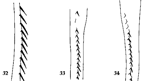
|
|
Figures 32-34. Ventral view of epipleuron showing where stridulatory ridge begins: 32) near outer edge of epipleuron (T. foveicollis); 33) at center of epipleuron (T. vittatus); 34) near inner edge of epipleuron (T. laticollis).
|
|
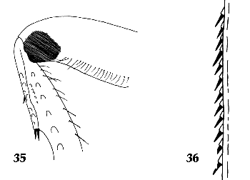
|
|
Figures 35-36. Stridulatory apparatus of Trizogeniates composed by: 35) stridulatory ridge (dorsal view of elytral margin); 36) stridulatory file (inner-lateral view of metafemur).
|
|
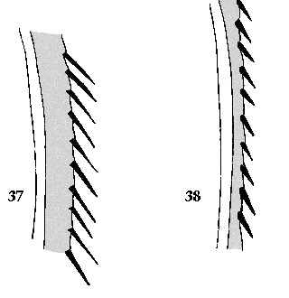
|
|
Figures 37-38. Ventral view of epipleuron showing width of inner epipleural region laterad of stridulatory ridge: 37) broad (T. dispar); 38) narrow (T. venezuelensis).
|
|
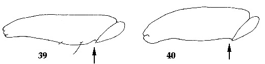
|
|
Figures 39-40. Ventral view of metafemur showing shape of posterior margin at base: 39) indented (T. bordoni); 40) not indented (T. montanus).
|
|
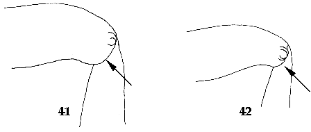
|
|
Figures 41-42. Ventral view of metafemur showing showing shape of posterior margin at apex: 41) extended and rounded (T. terricola); 42) weakly extended and weakly rounded (T. traubi).
|
|
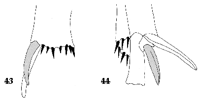
|
|
Figures 43-44. Male metatibial spurs showing shape of outer metatibal spur (shaded): 43) curved (outer lateral view, T. bicolor); 44) weakly curved (inner lateral view, T. foveicollis).
|
|
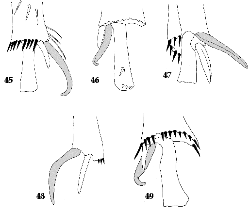
|
|
Figures 45-49. Male metatibial spurs showing shape of inner metatibial spur (shaded): 45) hooked (T. aphilus); 46) hooked (T. crispospinatus); 47) weakly curved (T. foveicollis); 48) strongly curved (T. schmidti); 49) hooked (T. tibialis).
|
|
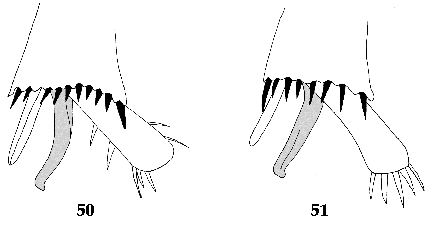
|
|
Figures 50-51. Female metatibial spurs showing shape of inner metatibial spur (shaded): 50) T. crispospinatus; 51) T. tibialis.
|
|
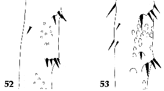
|
|
Figures 52-53. Surface of metatibia showing puncture shape: 52) punctures round (T. catoxanthus); 53) oval-shaped (T. montanus).
|
|
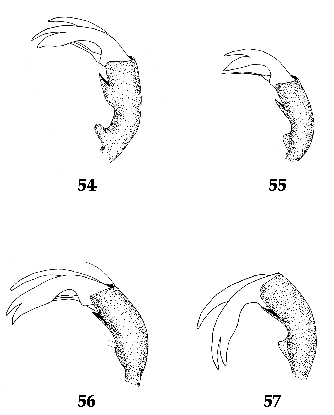
|
|
Figures 54-57. Male fifth metatarsomere showing presence and shape of protuberance: 54) protuberance rounded (T. dispar); 55) protuberance laterally flattened (T. ohausi); 56) protuberance absent (T. traubi); 57) protuberance laterally flattened (T. caiporae).
|
|
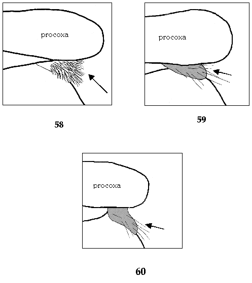
|
|
Figures 58-60. Lateral view showing shape of prosternal shield. 58)T. montanus, 59) T. traubi, 60) T. venezuelensis.
|
|
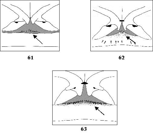
|
|
Figures 61-63. Ventral view of first sternite, showing shape of middle region. 61) simple (T. barrerai), 62) ventrally produced (T. caiporae), 63) ventrally produced (T. temporalis).
|
|
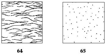
|
|
Figures 64-65. Surface of pygidium showing texture. 64) rugose (T. barrerai), 65) smooth (T. travassosi).
|
|

|
|
Figures 66-71. Caudal view of pygidium, showing shape of apical bead. 66) T. caiporae; 67) T. catsus; 68) T. crispospinatus, 69) T. laticollis, 70) T. geminatus, 71) T. trivittatus.
|
|

|
|
Figures 72-73. Lateral view of pygidium, showing shape. 72) T. travassosi; 73) T. geminatus.
|
|

|
|
Figures 74-76. Ventral view of last sternite of males, showing shape of emargination 74) T. aphilus; 75) T. crispospinatus; 76) T. tibialis.
|
|

|
|
Figures 77-82. Ventral view of last sternite of female, showing apical sculpture: 77) crenulate (T. bicolor); 78) shallowly bi-emarginate (T. geminatus); 79) shallowly emarginate (T. laticollis); 80) notched (T. tibialis); 81) deeply bi-emarginate(T. travassosi); 82) simple (T. trivittatus).
|
|

|
|
Figures 83-94. Caudal view of parameres of Trizogeniates species (all to same scale). 83) T. aphilus; 84) T. barrerai; 85) T. bicolor; 86) T. bordoni; 87) T. caiporae; 88) T. catoxanthus; 89) T. catsus; 90) T. cribicollis; 91) T. crispospinatus (form I); 92) T. crispospinatus (form II); 93) T. dispar; 94) T. foveicollis.
|
|

|
|
Figures 95-105. Caudal view (except where noted) of parameres of Trizogeniates species (all to same scale). 95) T. geminatus; 96) T. goyanus; 97) T. laticollis (form I); 98) T. laticollis (form II); 99) T. laticollis (frontal view, form III); 100) T. laticollis (lateral view, form I); 101) T. laticollis (lateral view, form II); 102) T. montanus; 103)T. ohausi; 104) T. planipennis; 105) T. schmidti.
|
|

|
|
Figures 106-113. Caudal view of parameres of Trizogeniates species (all to same scale). 106) T. temporalis; 107) T. terricola; 108) T. tibialis; 109) T. traubi; 110) T. travassosi; 111) T. trivittatus; 112) T. venezuelensis; 113) T. vittatus.
|
Trizogeniates calcaratus Ohaus
Trizogeniates calcaratus Ohaus, 1917: 40.
The type specimen of Trizogeniates calcaratus was not located in any collection, including ZMHB where the Ohaus types are housed. This species was described from one male specimen from Ecuador. The description is superficial and does not allow for a diagnosis. However, Ohaus (1917) mentioned that the specimen had the “upper metatibial spur bent or curved with its apex rounded and ventrally bent almost in a right angle”. This is similar to the species that have a hooked apex of the inner, metatibial spurs (T. tibialis, T. trivittatus, T. crispospinatus, T. aphilus). From the known species, no specimens from Ecuador show this character.
Trizogeniates costatus Ohaus
Trizogeniates costatus Ohaus, 1917: 43.
Type specimen was not found in any collection, including ZMHB where the Ohaus types are housed. Ohaus (1917) described this species from a male specimen collected in Brazil. The description is too general and the characters used are not diagnostic at the species level. Antonio Martínez and Kadlec identified several specimens as T. costatus, and these are conspecific with the type specimen of T. terricola (see the remarks for T. terricola). This suggests a possible synonymy, but it can’t be verified without the type material for T. costatus.
Trizogeniates laevis (Camerano)
Geniates laevis Camerano, 1878: 237.
Trizogeniates laevis (Camerano, 1878). New combination by Ohaus, 1922.
The type specimen was not located at any of the collections from where material was obtained or requested. Camerano described this species from a single male collected in Brazil. The description is brief and does not allow a diagnosis of the species.
Acknowledgments
I am indebted to Dr. Mary Liz Jameson and Dr. Brett Ratcliffe for critically reviewing the manuscript. Mary Liz Jameson, Aura Paucar, and Federico Ocampo are kindly acknowledged for testing and providing valuable comments on the key. I would like to thank the individuals and institutions from which specimens were borrowed: Manfred Uhlig, Bernard Jaeger, Joachim Schulze, and Hella Wendt (ZMHB), Karla Schneider and Joachim Händel (MLUH), and staff at DEI for their hospitality and assistance during my visits to their institutions. Dan Schmidt (Schuyler, NE) is gratefully acknowledged for the carbon dust habitus illustrations. I thank Chris Smith, Aura Paucar, Agustin Jiménez, and Federico Ocampo (all University of Nebraska) for their valuable support, help, and for providing me with literature, locality data, professional contacts, and type material from MACN. I thank Federico Ocampo for taking the photographs of the specimens and to Andrew Smith (UNSM) for providing professional contacts at other institutions.
This research was supported by an NSF/PEET grant (DEB-9712447) to B. C. Ratcliffe and M.L. Jameson. Partial support for the completion of this research was provided by the “Initiative in Ecology and Evolutionary Biology” of the School of Biological Sciences (University of Nebraska) and by the Department of Entomology (University of Nebraska).
References
Blackwelder RE. 1944. Checklist of the coleopterous insects of Mexico, Central America, the West Indies, and South America. Bull US Nat Mus 185, 189-341.
Burmeister HCC. 1844. Handbuch der Entomologie. Vol. 4 Pt. 1, Berlin.
Burmeister HCC. 1855. Handbuch der Entomologie. Vol. 4. Pt. 2, Berlin.
Camerano L. 1878. Studi intorno alle species del genere Geniates Kirby esistenti nel R. Museo Zoologico di Torino. Atti Accad Sc Torino 14, 229 -274.
Casey TL. 1915. A review of the American species of Rutelinae, Dynastinae, and Cetoniinae. Mem Coleop 6: 1-394.
[CZN] Commission on zoological Nomenclature. 1999. International Code of Zoological Nomenclature. 4th ed. The international trust for zoological nomenclature.
Evenhuis NL, Nishida GM, Samuelson GA. 2000. Insect and Spider Collections of the World Web Site. http://www.bishopmuseum.org/bishop/ento/codens-r-us.html>. Version: 7 March 2000. Accessed: September 2000.
Jameson ML. 1998 (1997). Phylogenetic analysis of the subtribe Rutelina and revision of the Rutela generic groups (Coleoptera: Scarabaeidae: Rutelinae: Rutelini). Bull Univ Nebraska St Mus 14:1-184.
Lucas PH. 1857. Animaux nouveaux ou rares recueillis pendant l’expédition dans les parties centrales de l’Amérique du Sud, de Rio de Janeiro a Lima, et de Lima au Para; exécutée par ordre du Government français pendant les années 1843 a 1847, sous la direction du Comte Francis de Castelnau. (Entomologie. Bertrand: Paris)
Machatschke J. 1965. Coleoptera Lamellicornia. Fam. Scarabaeidae, Subfam. Rutelinae, Section Rutelinae Orthochilidae. Genera Insect 199C:1-145.
Machatschke J. 1972. Scarabaeidae: Melolonthidae, Rutelinae. Coleop Cat Sup 66(1):1-361.
Machatschke J. 1974. Scarabaeidae: Melolonthidae, Rutelinae. Coleop Cat Sup 66(2):363-429.
Martinez A. 1958. Algunos Geniatini bolivianos nuevos o poco conocidos (Col. Scarabaeidae, Rutelinae). Studia Entomol 1:407-420.
Martinez A. 1965. Nuevas especies de Trizogeniates (Col. Scarab. Rutel. Geniatini). An Soc Cient Argentina 179:3-16.
Ohaus F. 1903. Verzeichnis der von Herrn Richard Haensch in Ecuador gesammelten Ruteliden (Coleoptera lamellicornia). Berliner Entomol Z 48:215 -447.
Ohaus F. 1917. Neue Geniatinen (Col. lamell. Rutelin.). Stettiner Entomol Zeitung 78:3-53.
Ohaus F. 1918. Neue Geniatinen II (Col. lamell. Rutelin.). Stettiner Entomol Zeitung 79, 350-365.
Ohaus F. 1922. Beitrag zur Kenntnis der Ruteliden (Col. Lamell.). Dtsch Entomol Z 19:323-331.
Ohaus F. 1931. Neue Geniatinen IV (Col.). Stettiner Entomol Z 92:227-259.
Wheeler QD, Platnick NI. 2000. The phylogenetic species concept. In : Wheeler QD, Meier R, editors. Species concepts and phylogenetic theory. New York: Columbia Univ. p. 55-69.
Villatoro KE, Jameson ML. 2000. Annotated catalog and key to the genera of the world Geniatini (Coleoptera: Scarabaeidae: Rutelinae) Web Site. <http://www-museum.unl.edu/research/entomology/genicatalog.htm.


